. 2017 Jun 20;85(7):e00075-17.
doi: 10.1128/IAI.00075-17. Print 2017 Jul.

Toll-Like Receptor 2 and Mincle Cooperatively Sense Corynebacterial Cell Wall Glycolipids

Affiliations

Toll-Like Receptor 2 and Mincle Cooperatively Sense Corynebacterial Cell Wall Glycolipids

Judith Schick et al. Infect Immun. .

Abstract

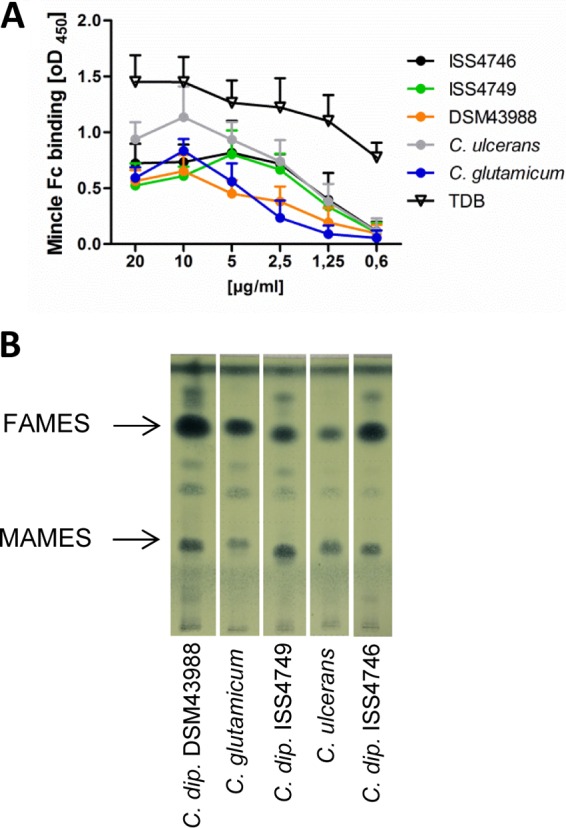
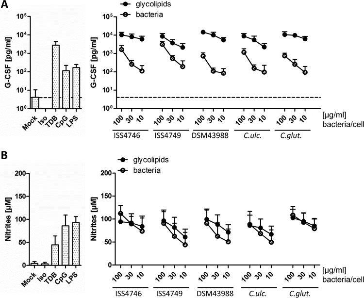
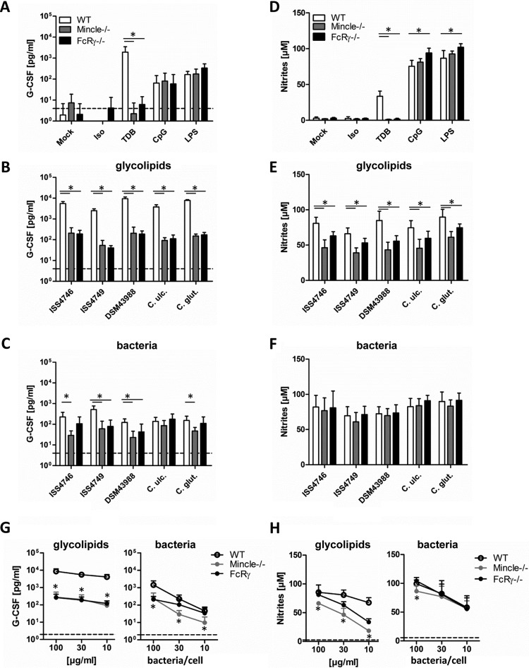
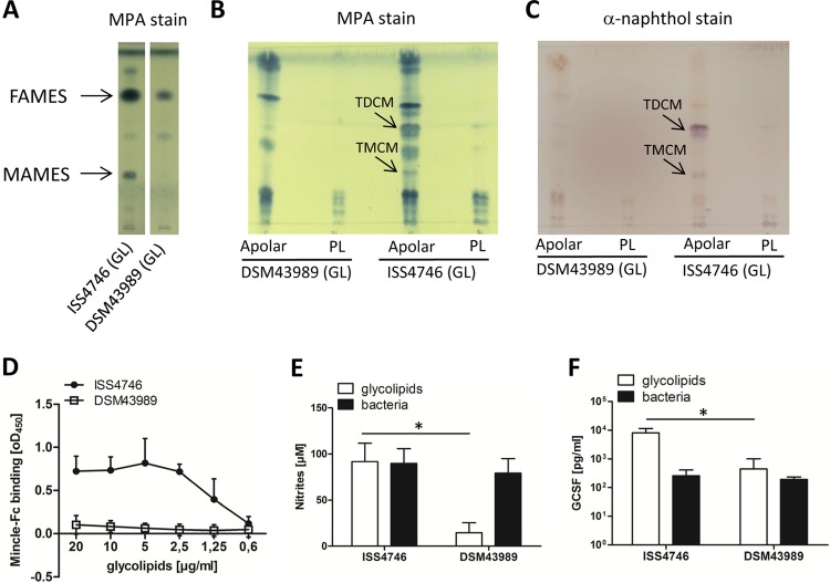
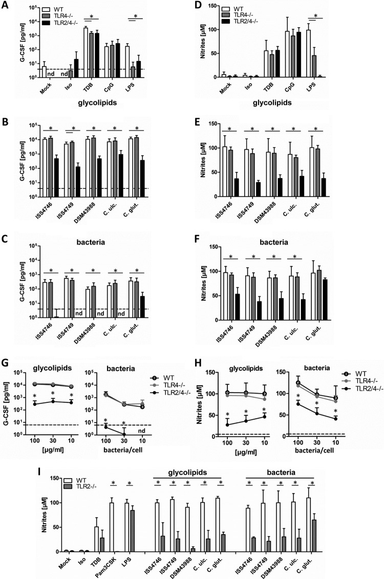
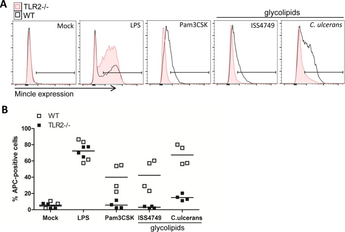
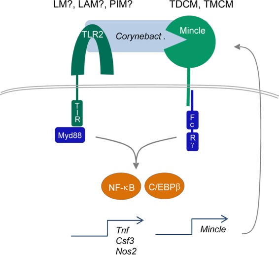
Nontoxigenic Corynebacterium diphtheriae and Corynebacterium ulcerans cause invasive disease in humans and animals. Host sensing of corynebacteria is largely uncharacterized, albeit the recognition of lipoglycans by Toll-like receptor 2 (TLR2) appears to be important for macrophage activation by corynebacteria. The members of the order Corynebacterineae (e.g., mycobacteria, nocardia, and rhodococci) share a glycolipid-rich cell wall dominated by mycolic acids (termed corynomycolic acids in corynebacteria). The mycolic acid-containing cord factor of mycobacteria, trehalose dimycolate, activates the C-type lectin receptor (CLR) Mincle. Here, we show that glycolipid extracts from the cell walls of several pathogenic and nonpathogenic Corynebacterium strains directly bound to recombinant Mincle in vitro Macrophages deficient in Mincle or its adapter protein Fc receptor gamma chain (FcRγ) produced severely reduced amounts of granulocyte colony-stimulating factor (G-CSF) and of nitric oxide (NO) upon challenge with corynebacterial glycolipids. Consistently, cell wall extracts of a particular C. diphtheriae strain (DSM43989) lacking mycolic acid esters neither bound Mincle nor activated macrophages. Furthermore, TLR2 but not TLR4 was critical for sensing of cell wall extracts and whole corynebacteria. The upregulation of Mincle expression upon encountering corynebacteria required TLR2. Thus, macrophage activation by the corynebacterial cell wall relies on TLR2-driven robust Mincle expression and the cooperative action of both receptors.

Keywords: C-type lectin receptor; Corynebacterium diphtheriae; Mincle; Toll-like receptor; cell wall lipids; corynomycolate; macrophage; mycolate; mycolic acid.


Figures

FIG 1
FIG 1
Cell wall extracts of corynebacteria bind the Mincle-Fc fusion protein in vitro. (A) The binding affinity of the Mincle-Fc fusion protein for different concentrations of corynebacterial glycolipid extracts was analyzed by using a Mincle-Fc binding assay. Data are depicted as mean values and standard deviations from 3 experiments performed in duplicates (n = 6). (B) TLC of fatty acid and mycolic acid methyl esters stained with MPA. C. dip., C. diphtheriae.
FIG 2
FIG 2
Corynebacterial glycolipids induce G-CSF and NO production in macrophages in a concentration-dependent manner. BMM from C57BL/6 mice were stimulated with different concentrations of glycolipids (100, 30, or 10 μg/ml) or heat-killed bacteria (100, 30, or 10 bacteria per cell), followed by determination of G-CSF (A) and nitrite (B) levels in cell culture supernatants via an ELISA and a Griess assay, respectively. The detection limit for G-CSF was 4 pg/ml (dotted line). For comparison, unstimulated cells or TDB-, CpG-, or LPS-stimulated cells were used as controls. Data are represented as means and standard deviations of results from 6 independent experiments performed in duplicates (n = 12). Iso, isopropanol; C. ulc., C. ulcerans; C. glut., C. glutamicum.
FIG 3
FIG 3
Mincle/FcRγ deficiency impairs G-CSF and NO production in macrophages stimulated with corynebacteria or their cell wall glycolipids. (A to C) BMM from Mincle- or FcRγ-deficient mice and their respective wild-type controls were stimulated with different glycolipid extracts (30 μg/ml) or the corresponding heat-killed bacteria (30 bacteria per cell) as well as controls for 48 h, as indicated. G-CSF cytokine levels in cell culture supernatants were determined by using an ELISA. The detection limit was 4 pg/ml (dotted line). (D to F) NO production in cell culture supernatants was determined by using a Griess assay. BMM were cotreated with 10 ng/ml IFN-γ. (G and H) BMM were stimulated with different concentrations of glycolipids (100, 30, or 10 μg/ml) or heat-killed bacteria (100, 30, or 10 bacteria per cell). Data shown were obtained with glycolipids from C. diphtheriae ISS4746; extracts from the other corynebacterial strains/species showed similar dose-response curves (not shown for clarity). G-CSF and NO production was analyzed as described above. Dotted lines represent G-CSF or nitrite levels from unstimulated BMM. Note that data points for some Mincle−/− and FcRγ−/− BMM overlap in panel G. Data are depicted as means and standard deviations of results from 3 independent experiments performed in duplicates (n = 6). In panels B to H, * indicates a P value of <0.05 for the indicated comparison between genotypes.
FIG 4
FIG 4
Cell wall extracts of C. diphtheriae DSM43989 fail to activate macrophages and do not bind Mincle-Fc. (A) TLC of different lipid extracts stained by using MPA. (B and C) Apolar compounds and phospholipids (PL) of glycolipid (GL) extracts were purified and also detected by TLC using MPA (B) or α-naphthol (C). (D) The binding affinity of the Mincle-Fc fusion protein for different concentrations of C. diphtheriae ISS4746 and DSM43989 glycolipid extracts was analyzed by using a Mincle-Fc binding assay. Shown are means and standard deviations of results from three independent experiments performed in duplicates (n = 6). (E and F) Bone marrow-derived murine macrophages from C57BL/6 mice were stimulated with lipid extracts (30 μg/ml) or the corresponding heat-killed bacteria (30 bacteria per cell) from C. diphtheriae ISS4746 or DSM43989 in the presence (E) or absence (F) of 10 ng/ml IFN-γ. NO production (E) and G-CSF levels (F) were measured in the supernatant 48 h after stimulation. Data are depicted as means and standard deviations of results from 6 independent experiments performed in duplicates (n = 12).
FIG 5
FIG 5
TLR2 deficiency impairs G-CSF and NO production in macrophages stimulated with corynebacteria or their cell wall glycolipids. (A to C) BMM from TLR4-deficient or TLR2/4-double-deficient mice and their respective wild-type controls were stimulated with different glycolipid extracts (30 μg/ml) or the corresponding heat-killed bacteria (30 bacteria per cell) as well as controls for 48 h, as indicated. G-CSF cytokine levels in cell culture supernatants were determined by using an ELISA. The detection limit was 4 pg/ml (dotted line). nd, not detectable. (D to F) NO production in cell culture supernatants was determined by using a Griess assay. BMM were cotreated with 10 ng/ml IFN-γ. BMM were stimulated with different concentrations of glycolipids (100, 30, or 10 μg/ml) or heat-killed bacteria (100, 30, or 10 bacteria per cell). Data shown were obtained with glycolipids from C. diphtheriae ISS4746; extracts from the other corynebacterial strains/species showed similar dose-response curves (not shown for clarity). (G and H) G-CSF levels and NO production were analyzed as described above. Dotted lines represent G-CSF or nitrite levels from unstimulated BMM. Note that data points for WT and TLR4−/− BMM overlap in panel G. Data are depicted as means and standard deviations of results from 3 independent experiments performed in duplicates (n = 6). (I) Wild-type and TLR2-deficient BMM were stimulated as described above for panels A to C, followed by NO measurement. Data are depicted as means and standard deviations of results from 3 independent experiments performed in duplicates (n = 6). In panels B, C, and E to I, * indicates a P value of <0.05.
FIG 6
FIG 6
TLR2 deficiency leads to impaired Mincle upregulation on the cell surface. Shown are data from flow cytometric analysis of Mincle receptor expression on TLR2-deficient BMM and the respective wild-type controls. BMM were stimulated with 30 μg/ml cell wall extract of C. diphtheriae ISS4749 or C. ulcerans for 48 h. Unstimulated cells (mock) or BMM stimulated with 10 ng/ml LPS or 50 ng/ml Pam3CSK4 were used as controls. Panel A depicts representative histograms, indicating the gate used for the determination of Mincle-expressing macrophages shown in panel B. Data are depicted as single values from 2 independent experiments with biological duplicates (n = 4).
FIG 7
FIG 7
Synergistic induction of macrophage activation by the corynebacterial cell wall through TLR2 and Mincle signaling. The cell wall of corynebacteria contains TLR2 ligands, which activate macrophages through MyD88-dependent signaling. Lipomannan (LM) and Lipoarabinomannan (LAM) have been identified as TLR2 ligands from corynebacteria (31); PIMs from mycobacteria activate TLR2 (44) and are present in corynebacterial glycolipid extracts. Mincle binds to glycolipids containing mycolates (likely TDCM and TMCM), which activates FcRγ-Syk-dependent gene expression (Csf3 encodes G-CSF, and Nos2 encodes iNOS). Both pathways synergize at the level of NF-κB. In addition, TLR2- and C/EBPβ-dependent upregulation of Mincle mRNA and protein expression increases cell surface Mincle receptor expression and responsiveness to TDCM/TMCM in murine macrophages.

References

    1. Wong TP, Groman N. 1984. Production of diphtheria toxin by selected isolates of Corynebacterium ulcerans and Corynebacterium pseudotuberculosis. Infect Immun 43:1114–1116. - PMC - PubMed
    1. Dorella FA, Pacheco LG, Oliveira SC, Miyoshi A, Azevedo V. 2006. Corynebacterium pseudotuberculosis: microbiology, biochemical properties, pathogenesis and molecular studies of virulence. Vet Res 37:201–218. doi: 10.1051/vetres:2005056. - DOI - PubMed
    1. Hacker E, Antunes CA, Mattos-Guaraldi AL, Burkovski A, Tauch A. 2016. Corynebacterium ulcerans, an emerging human pathogen. Future Microbiol 11:1191–1208. doi: 10.2217/fmb-2016-0085. - DOI - PubMed
    1. Zakikhany K, Neal S, Efstratiou A. 2014. Emergence and molecular characterisation of non-toxigenic tox gene-bearing Corynebacterium diphtheriae biovar mitis in the United Kingdom, 2003-2012. Euro Surveill 19(22):pii=20819. doi: 10.2807/1560-7917.ES2014.19.22.20819. - DOI - PubMed
    1. Zasada AA. 2013. Nontoxigenic highly pathogenic clone of Corynebacterium diphtheriae, Poland, 2004-2012. Emerg Infect Dis 19:1870–1872. doi: 10.3201/eid1911.130297. - DOI - PMC - PubMed

Publication types

LinkOut - more resources