. 2017 Jun 29;37(14):e00659-16.
doi: 10.1128/MCB.00659-16. Print 2017 Jul 15.

Progerin-Induced Replication Stress Facilitates Premature Senescence in Hutchinson-Gilford Progeria Syndrome

Affiliations

Progerin-Induced Replication Stress Facilitates Premature Senescence in Hutchinson-Gilford Progeria Syndrome

Keith Wheaton et al. Mol Cell Biol. .

Abstract

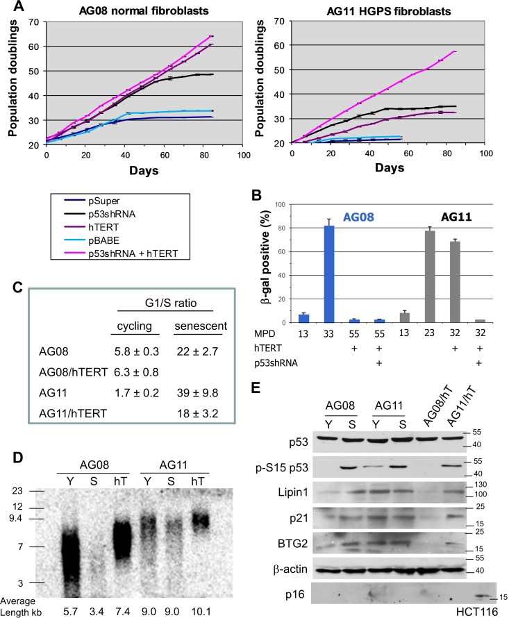
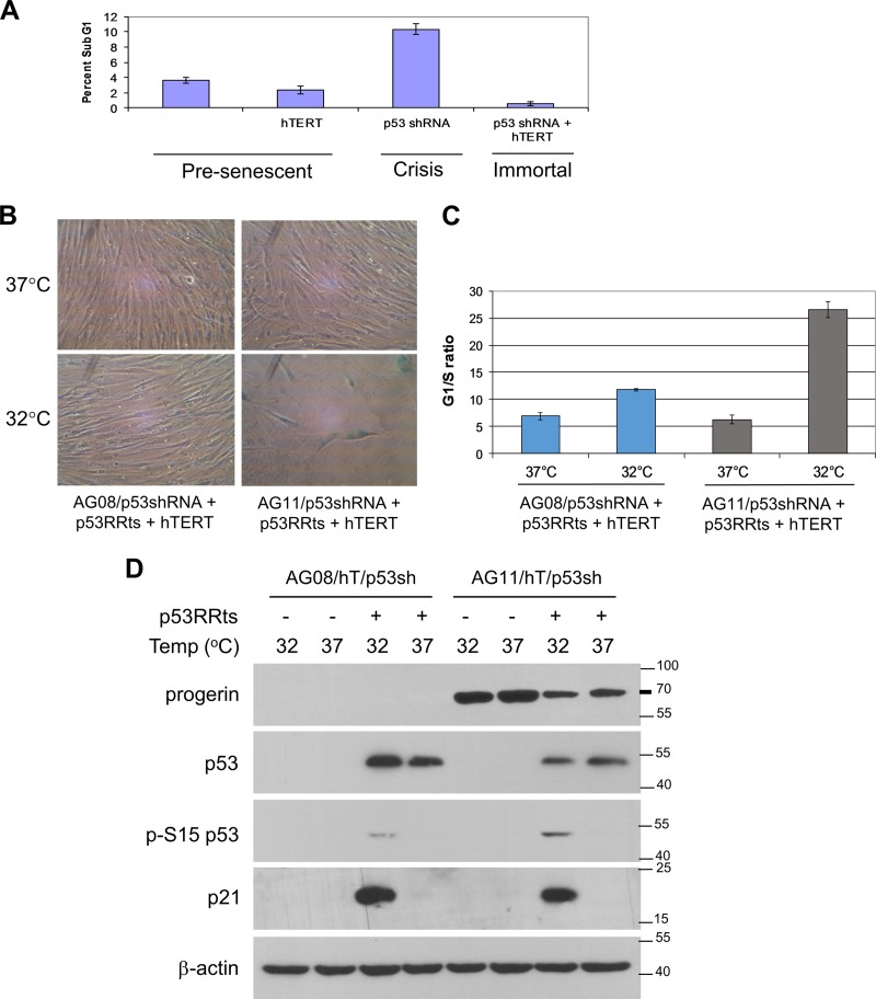
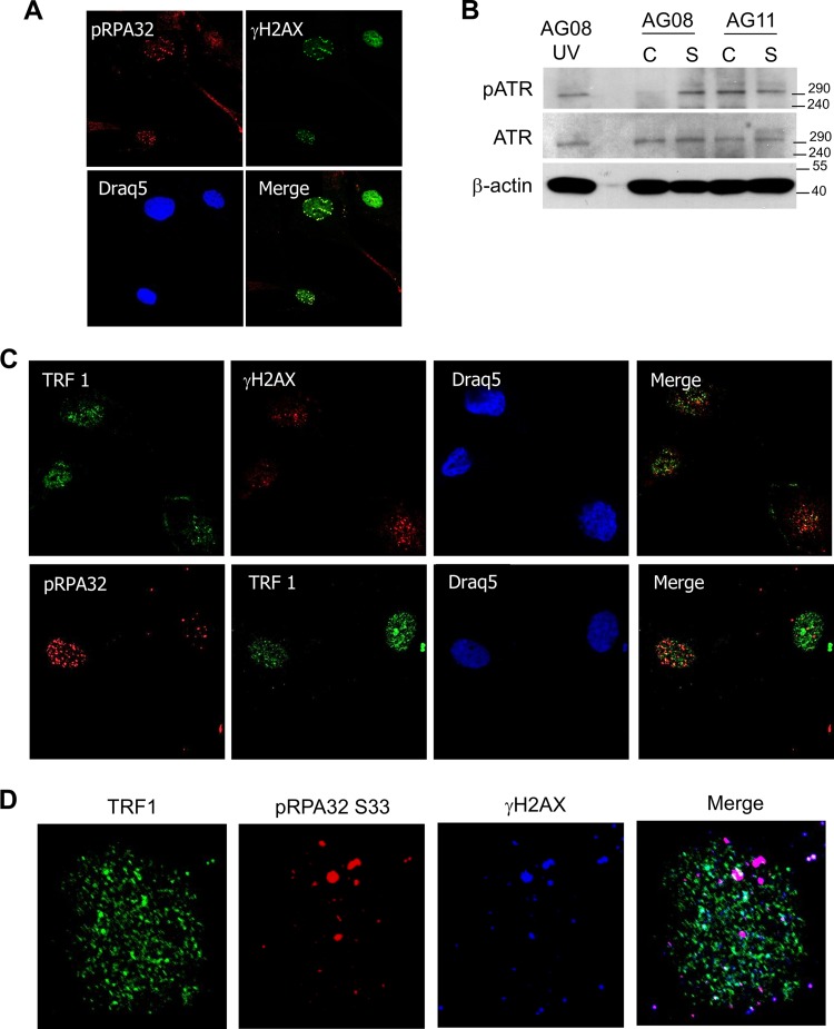
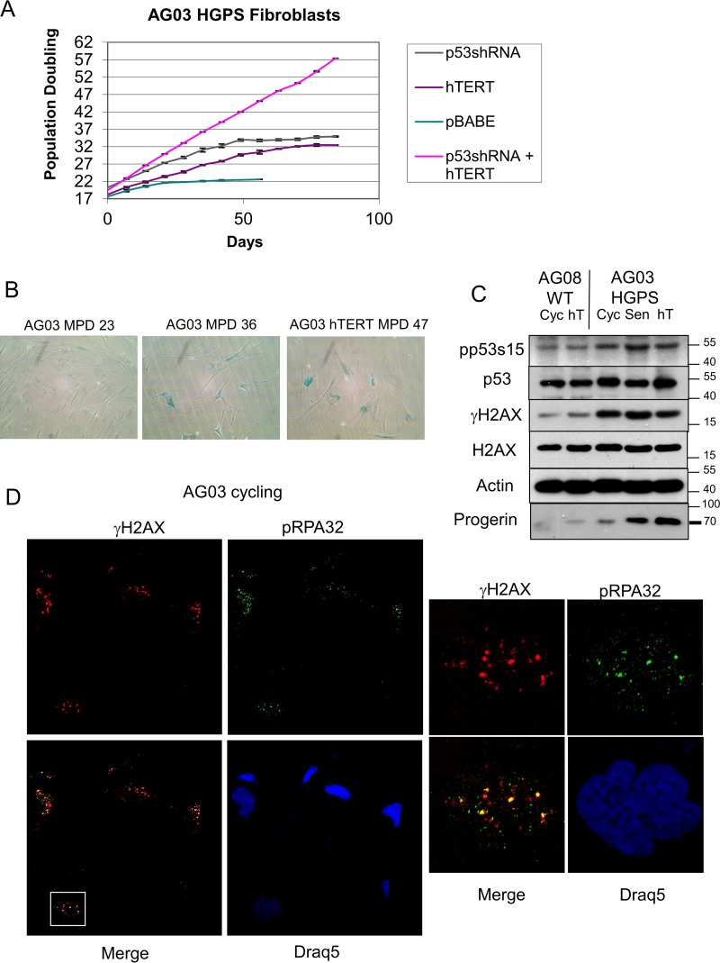
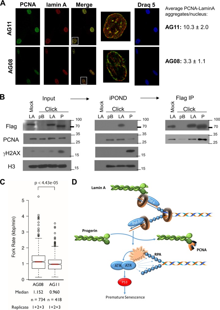
Hutchinson-Gilford progeria syndrome (HGPS) is caused by a mutation in LMNA that produces an aberrant lamin A protein, progerin. The accumulation of progerin in HGPS cells leads to an aberrant nuclear morphology, genetic instability, and p53-dependent premature senescence. How p53 is activated in response to progerin production is unknown. Here we show that young cycling HGPS fibroblasts exhibit chronic DNA damage, primarily in S phase, as well as delayed replication fork progression. We demonstrate that progerin binds to PCNA, altering its distribution away from replicating DNA in HGPS cells, leading to γH2AX formation, ATR activation, and RPA Ser33 phosphorylation. Unlike normal human cells that can be immortalized by enforced expression of telomerase alone, immortalization of HGPS cells requires telomerase expression and p53 repression. In addition, we show that the DNA damage response in HGPS cells does not originate from eroded telomeres. Together, these results establish that progerin interferes with the coordination of essential DNA replication factors, causing replication stress, and is the primary signal for p53 activation leading to premature senescence in HGPS. Furthermore, this damage response is shown to be independent of progerin farnesylation, implying that unprocessed lamin A alone causes replication stress.

Keywords: HGPS; aging; p53; progerin; senescence; telomere.


Figures

FIG 1
FIG 1
Premature senescence in HGPS cells is dependent on p53 and not dependent on telomere shortening. (A) Measurement of the number of mean population doublings over the life span of normal AG08 fibroblasts (left) and HGPS AG11 fibroblasts (right) expressing p53 shRNA, hTERT, or p53shRNA plus hTERT. Data represent averages from 3 biological replicates counted with 4 technical replicates each week. Standard error of the mean (SEM) values are shown as error bars. (B) β-Galactosidase staining of normal AG08 and HGPS AG11 fibroblasts expressing hTERT alone or p53shRNA plus hTERT at the indicated mean population doubling (MPD). (C) The G1/S ratios were determined from cell cycle profiles obtained by flow cytometry after propidium iodide staining. Cellular senescence is reflected by an increase in the G1/S ratio. (D) TRF assay of AG08 and AG11 cells showing overall telomere length in young (Y), senescent (S), and hTERT (hT)-expressing cells. This assay was repeated 3 times, and a representative result is shown. The average telomere length, determined by pixel density maxima, is shown. (E) Western blot analysis of young cycling and senescent AG08 and AG11 cells; AGO8 and AG11 cells expressing hTERT at population doubling 35 were also compared. Protein extracts were immunoblotted with the indicated antibodies. HCT116 lysates served as a positive control for p16 expression.
FIG 2
FIG 2
Progerin-induced premature senescence is dependent on p53. (A) Determination of crisis (apoptosis) in AG11 HGPS cells expressing pBABE vector control (MPD 25), hTERT (MPD 45), p53 shRNA (MPD 49), or both hTERT and p53shRNA (MPD 45). Apoptosis was determined by flow cytometry on the basis of sub-G1 content after propidium iodide staining. At the time of analysis, MPD 49 p53 shRNA cells were in a state of decline in which overall cell numbers were decreasing, while all other cell lines were actively growing. (B to D) p53RRts and hTERT were coexpressed in normal AG08/p53 shRNA and HGPS AG11/p53 shRNA. The cells were assessed at an MPD of 60 (AG08/p53sh/p53RRts/hTERT) or MPD of 50 (AG11/p53sh/p53RRts/hTERT). hTERT was expressed so that the contribution of progerin could be assessed independently of telomere erosion during premature senescence. (B) Cell growth was assessed at 37°C and at 32°C. Cells were visualized after β-galactosidase staining. hTERT-p5RRts-expressing AG11 cells at 32°C were 82% ± 5.6% positive β-galactosidase, demonstrating telomere-independent senescence. (C) The cells used for panel B were stained with propidium iodide, and the G1/S ratios were obtained from the cell cycle profiles using flow cytometry. Values shown represent the means and SEM (n = 3). (D) Western blot analysis confirming p53 knockdown and p53RRts and progerin expression in the cells used for panel B. The activation of p53 at 32°C is demonstrated by the upregulation of p21 and by the phosphorylation of p53 at Ser15.
FIG 3
FIG 3
Cycling HGPS cells exhibit chronic DNA damage associated with DNA replication stress. (A) Immunostaining for γH2AX and 53BP1 in cycling or noncycling (serum-starved) HGPS AG11 or cycling-normal AG08 cells. Draq5 was used to stain nuclei. (B) Western blot analysis of γH2AX, H2AX, p53, phospho-Ser15 p53, and p21 in cycling (Cyc) and serum-starved (SS) AG08 and AG11 cells. Cells were serum starved for 5 days. (C) Cells were labeled with BrdU (10 nM) for 8 h and immunostained with antibodies against 53BP1. The proportion of BrdU-positive and BrdU-negative cells showing ≥5 53BP1 foci is indicated by the black shading. Data presented are means and SEM from 3 independent experiments; 150 to 200 cells were counted in each experiment. (D) Young cycling AG08 (MPD 22) and AG11 (MPD 13) cells were subjected to cell cycle analysis by flow cytometry after propidium iodide staining. Also shown are hTERT-expressing AG08 and AG11 cells. The following percentages of cells were in S phase: AG08, 13% ± 0.7%; AG11, 35% ± 2.7%; AG08 hT, 12.5% ± 1.4%; and AG11, 18.6% ± 1%. Shown are the mean values and SEM (n = 3). (E) The proportion of cells in different phases of the cell cycle that were expressing γH2AX was measured by flow cytometry using propidium iodide to determine DNA content and antibodies to γH2AX to determine the extent of DNA damage. The values shown represent the means and SEM from 3 independent experiments.
FIG 4
FIG 4
DNA damage from replication stress in HGPS cells colocalizes with pRPA32 and not with telomeres (TRFI). (A) Immunostaining for γH2AX and pRPA32 in cycling AG11 cells. Phospho-RPA32 (Ser33) serves as a marker for single-stranded DNA that is associated with stalled DNA replication forks. (B) Western blot analysis of phospho-ATR and total ATR in cycling (C) and senescent (S) AG08 and AG11 cells. β-Actin served as a loading control. UV-treated normal cells (AG08) served as a positive control for pATR expression. (C) Immunostaining for γH2AX, TRF1, and pRPA32 in AG11 HGPS cells. pRPA is a marker for single-stranded DNA that accumulates at stalled DNA replication forks, and TRF1 is a marker for telomeres. AG11 cells were doubly stained with TRF1 and γH2AX or pRPA32 and TRF2. (D) Nucleus of an AG11 cell that was triply stained with pRPA (Dylight 649), TRF1 (Alexa Fluor 488), and γH2AX (Cy3).
FIG 5
FIG 5
AG03 HGPS fibroblasts show p53-dependent and telomere-independent premature senescence and replication stress. (A) Measurement of the number of mean population doublings over the life span of HGPS AG03 fibroblasts expressing pBABE vector control, p53 shRNA, hTERT, or p53shRNA plus hTERT. Data represent averages from 3 biological replicates counted with 4 technical replicates each week. SEM values are shown as error bars. (B) β-Galactosidase staining of cycling low-passage-number HGPS AG03 cells (MPD 23), senescent high-passage-number AG03 cells (MPD 36), and senescent AG03/hTERT cells (MPD 47). (C) Western blot analysis of young cycling normal AG08 and HGPS AG03 cells, immortal AG08/hTERT cells at MPD 44, and senescent AG03/hTERT cells at MPD 46. Protein extracts were immunoblotted with the indicated antibodies. Progerin levels, as expected, accumulate with population doubling. (D) Immunostaining for γH2AX (Cy3) and pRPA32 (Alexa Fluor 488) in cycling low-passage-number HGPS AG03 cells. Phospho-RPA32 (Ser33) serves as a marker for single-stranded DNA that is associated with stalled DNA replication forks. Draq5 was used to stain nuclei. The white box indicates the magnified view of a single nucleus to demonstrate colocalization of γH2AX with pRPA32.
FIG 6
FIG 6
Progerin sequesters PCNA. (A) Immunostaining for lamin A (Cy3) and PCNA (Alexa Fluor 488) in normal AG08 and HGPS AG11 cells. The white boxes indicate the magnified view of a single nucleus to demonstrate colocalization and PCNA aggregation. These aggregates were quantitated by analysis of the maxima of merged images using ImageJ. Numbers represent the averages from 100 to 200 cells of each type from two biological replicates. (B) The iPOND procedure was carried out in BJ cells (MPD 58-60) expressing vector control (pB), Flag-lamin A (LA), or Flag-progerin (P). The click reaction catalyzes the addition of biotin-azide to EdU-labeled DNA. A mock reaction without biotin-azide was carried out on lamin A input sample as a control. Antistreptavidin beads were used to pull down biotin-labeled chromatin and associated proteins. (Middle) This was followed by immunoblotting to detect PCNA, Flag, γH2AX, and H3 histone. (Right) After iPOND, the depleted extracts were subjected to immunoprecipitation (IP) with FLAG antibody, followed by Western blotting with FLAG or PCNA antibodies. (C) The rates of DNA replication fork progression in normal AG08 cells and HGPS AG11 cells (both MPD of 19) were measured using DNA combing. The distributions of rates from three replicates are presented as a box plot where the median is indicated by the red horizontal bar, the box spans the first through third quartiles, the whiskers extend to the last data points within 1.5 times the interquartile range, and outliers are plotted as circles. The distributions were compared using the Mann-Whitney U test, and the P value is indicated. (D) Accumulation of progerin leads to PCNA aggregation, replication stress, and p53 activation. The data are consistent with a model in which the accumulation of progerin causes the sequestration of PCNA, leading to its aggregation away from replication. The absence and miscoordination of PCNA leads to replication stress and the subsequent activation of p53. p53 activation in turn leads to premature senescence in HGPS cells.
FIG 7
FIG 7
Progerin and progerin C661M promote premature senescence and replication stress. (A and B) Measurement of the number of mean population doublings in normal BJ fibroblasts (A) or normal AG08 fibroblasts (B) expressing progerin, progerin C661M, hTERT, p53 shRNA, or empty vector (pBABE) as indicated. Data represent averages from 3 biological replicates counted with 4 technical replicates each week. SEM values are shown as error bars. Senescent arrest was confirmed by acidic β-galactosidase, with 80% ± 4.4% in progerin and 82% ± 3.9% in progerin C661M staining blue. (C) Western blot analysis of BJ and BJ/hTERT cells expressing empty vector (pBABE), progerin, or progerin C661M. Protein extracts were immunoblotted with the indicated antibodies. (D) Western blot analysis of AG08 and AG08/hTERT cells expressing empty vector (pBABE), progerin, or progerin C661M. Protein extracts were immunoblotted with the indicated antibodies. A, processed lamin A; P C611M, progerin C611M; P, progerin. Note that the progerin antibody used on the Western blot does not detect the progerin C611M mutant. p53 activation is reflected by phospho-Ser15 p53 and p21 expression. β-Actin served as a loading control. HCT116 cells served as a positive control for p16 expression.
FIG 8
FIG 8
Accumulation of progerin induces DNA damage and replication stress. (A) Immunostaining of γH2AX, 53BP1, and pRPA32 in normal AG08 fibroblasts expressing ectopic progerin. (B) Immunostaining of γH2AX, 53BP1, and pRPA32 in normal AG08 fibroblasts expressing ectopic progerin C661M. (C) Immunostaining using antiprogerin (Cy3-secondary antibody) and anti-53BP1 (Alexa Fluor 488-secondary antibody) and Draq5 in HGPS AG11 cells. Arrows indicate cells that lack endogenous progerin staining. (D) Cell counts of cells depicted in panel C showing the percentage of AG11 HGPS cells that immunostain (+) for progerin (red bars) with increasing mean population doublings (MPD). The proportion of cells with 5 or more 53BP1 foci is also shown. Data represent 100 to 200 cells in 3 individual experiments.

Similar articles

Cited by

References

    1. Eriksson M, Brown WT, Gordon LB, Glynn MW, Singer J, Scott L, Erdos MR, Robbins CM, Moses TY, Berglund P, Dutra A, Pak E, Durkin S, Csoka AB, Boehnke M, Glover TW, Collins FS. 2003. Recurrent de novo point mutations in lamin A cause Hutchinson-Gilford progeria syndrome. Nature 423:293–298. doi:10.1038/nature01629. - DOI - PMC - PubMed
    1. Burtner CR, Kennedy BK. 2010. Progeria syndromes and ageing: what is the connection? Nat Rev Mol Cell Biol 11:567–578. doi:10.1038/nrm2944. - DOI - PubMed
    1. Liu B, Wang J, Chan KM, Tjia WM, Deng W, Guan X, Huang JD, Li KM, Chau PY, Chen DJ, Pei D, Pendas AM, Cadinanos J, Lopez-Otin C, Tse HF, Hutchison C, Chen J, Cao Y, Cheah KS, Tryggvason K, Zhou Z. 2005. Genomic instability in laminopathy-based premature aging. Nat Med 11:780–785. doi:10.1038/nm1266. - DOI - PubMed
    1. Liu Y, Rusinol A, Sinensky M, Wang Y, Zou Y. 2006. DNA damage responses in progeroid syndromes arise from defective maturation of prelamin A. J Cell Sci 119:4644–4649. doi:10.1242/jcs.03263. - DOI - PMC - PubMed
    1. Kennedy BK, Barbie DA, Classon M, Dyson N, Harlow E. 2000. Nuclear organization of DNA replication in primary mammalian cells. Genes Dev 14:2855–2868. doi:10.1101/gad.842600. - DOI - PMC - PubMed

Publication types

Grants and funding

LinkOut - more resources