. 2017 Jun 29;37(14):e00075-17.
doi: 10.1128/MCB.00075-17. Print 2017 Jul 15.

An In Vitro TORC1 Kinase Assay That Recapitulates the Gtr-Independent Glutamine-Responsive TORC1 Activation Mechanism on Yeast Vacuoles

Affiliations

An In Vitro TORC1 Kinase Assay That Recapitulates the Gtr-Independent Glutamine-Responsive TORC1 Activation Mechanism on Yeast Vacuoles

Mirai Tanigawa et al. Mol Cell Biol. .

Abstract

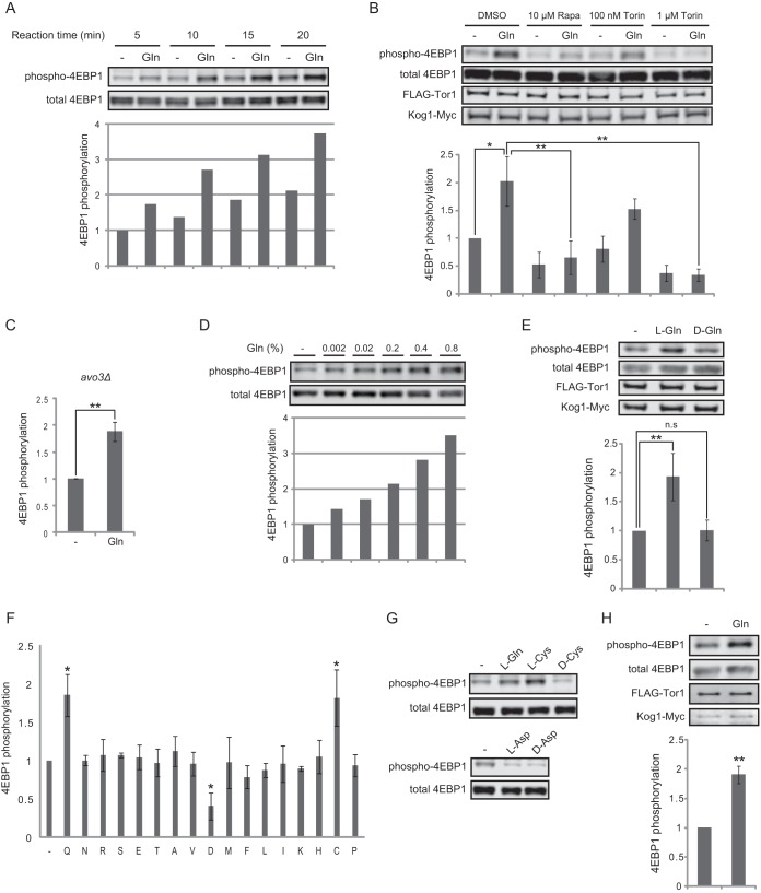
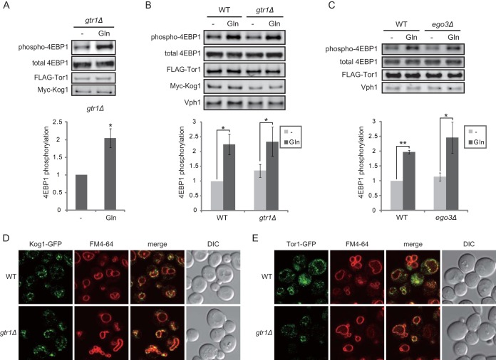
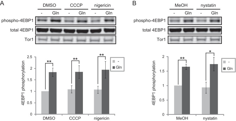
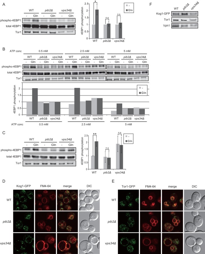
Evolutionarily conserved target of rapamycin (TOR) complex 1 (TORC1) responds to nutrients, especially amino acids, to promote cell growth. In the yeast Saccharomyces cerevisiae, various nitrogen sources activate TORC1 with different efficiencies, although the mechanism remains elusive. Leucine, and perhaps other amino acids, was reported to activate TORC1 via the heterodimeric small GTPases Gtr1-Gtr2, the orthologues of the mammalian Rag GTPases. More recently, an alternative Gtr-independent TORC1 activation mechanism that may respond to glutamine was reported, although its molecular mechanism is not clear. In studying the nutrient-responsive TORC1 activation mechanism, the lack of an in vitro assay hinders associating particular nutrient compounds with the TORC1 activation status, whereas no in vitro assay that shows nutrient responsiveness has been reported. In this study, we have developed a new in vitro TORC1 kinase assay that reproduces, for the first time, the nutrient-responsive TORC1 activation. This in vitro TORC1 assay recapitulates the previously predicted Gtr-independent glutamine-responsive TORC1 activation mechanism. Using this system, we found that this mechanism specifically responds to l-glutamine, resides on the vacuolar membranes, and involves a previously uncharacterized Vps34-Vps15 phosphatidylinositol (PI) 3-kinase complex and the PI-3-phosphate [PI(3)P]-binding FYVE domain-containing vacuolar protein Pib2. Thus, this system was proved to be useful for dissecting the glutamine-responsive TORC1 activation mechanism.

Keywords: Saccharomyces cerevisiae; TOR kinase; TORC1; Vps34; glutamine; in vitro kinase assay.


Figures

FIG 1
FIG 1
The newly developed in vitro TORC1 kinase assay. (A) Time course of 4EBP1 phosphorylation in the in vitro TORC1 kinase assay. Semi-intact cells prepared from wild-type (TS275) cells were used in the in vitro kinase assay using 4EBP1 as the substrate. The reactions were started by adding ATP with l-glutamine (Gln) (final concentration, 0.2%) or without l-glutamine (−), and the reaction mixtures were incubated at 30°C for the indicated periods. The phosphorylation status of 4EBP1 Thr37/46 was detected by Western blotting with an anti-phospho-T37/46-4EBP1-specific antibody. (B) l-Glutamine-responsive TORC1 activation in vitro. An in vitro kinase assay using semi-intact cells was performed as described above for panel A, except that the reaction mixture was incubated for 10 min. The reaction was performed with or without the indicated amount of rapamycin (Rapa) or torin-1 (Torin). FLAG-tagged Tor1 and Myc-tagged Kog1 were detected as loading controls. The bar graph shows the mean ratios of phosphorylated 4EBP1 to total 4EBP1, normalized to the values for the dimethyl sulfoxide (DMSO)-treated samples to which glutamine was not added (mean ± standard deviation [SD] [error bar]; n = 4). Mean values that are significantly different by the Student t test are indicated by bars and asterisks as follows: *, P < 0.05; **, P < 0.01. (C) TORC2 does not contribute to glutamine-responsive 4EBP1 phosphorylation. Semi-intact cells were prepared from avo3Δ (MH980) cells harboring a YPK2(D239A)-encoding multicopy plasmid [YEp352-YPK2(D239A)-HA], and an in vitro kinase assay was performed as described above for panel B (mean ± SD; n = 3). **, P < 0.01. (D) Glutamine activates TORC1 in vitro in a dose-dependent manner. An in vitro kinase assay was performed as described above for panel B in the presence of the indicated concentrations of l-glutamine. (E) d-Glutamine does not activate TORC1 in vitro. l-Glutamine or d-glutamine (final concentration, 0.2%) was added to the kinase reaction performed as described above for panel B. The bar graph shows the quantification of phosphorylated 4EBP1 (mean ± SD; n = 4). **, P < 0.01; n.s., not significant. (F) Glutamine and cysteine activate TORC1 in vitro. The indicated l-amino acids (final concentration, 0.2%) were added to the kinase reactions performed as described above for panel B. The bar graph shows the quantification of phosphorylated 4EBP1 (mean ± SD; n = 3). *, P < 0.05. (G) In vitro responsiveness of TORC1 activity to cysteine is stereospecific, but that to aspartic acid is not. An in vitro kinase assay using semi-intact cells prepared from wild-type (TM141) cells was performed as described above for panel B in the presence of the indicated amino acids (final concentration, 0.2%). (H) TORC1 in purified vacuoles exhibits glutamine-responsive activation. Vacuoles were isolated from wild-type (TS270) cells, and the in vitro kinase assay was performed as described above for panel B, except that purified vacuoles were used instead of semi-intact cells. The bar graph shows the quantification of phosphorylated 4EBP1, normalized to the values for the samples to which glutamine was not added (mean ± SD; n = 4). **, P < 0.01.
FIG 2
FIG 2
The EGO complex is dispensable for glutamine-responsive TORC1 activation in vitro. (A) Gtr1 is not required for glutamine responsiveness of semi-intact cells. An in vitro kinase assay was performed using semi-intact cells prepared from gtr1Δ (MH974) cells. FLAG-tagged Tor1 and Myc-tagged Kog1 were detected as loading controls. The bar graph shows the mean ratio of phosphorylated 4EBP1 to total 4EBP1, normalized to the values for the samples to which glutamine was not added (mean ± SD; n = 4). *, P < 0.05. (B) Gtr1 is not required for glutamine responsiveness of vacuoles. An in vitro kinase assay was performed using vacuoles isolated from wild-type (WT) (TS270) or gtr1Δ (MH974) cells. FLAG-tagged Tor1 (FLAG-Tor1), Myc-tagged Kog1, and Vph1 (vacuolar marker) were detected as loading controls. The bar graph shows the mean ratios of phosphorylated 4EBP1 to total 4EBP1/Tor1, normalized to the values for the wild-type samples to which glutamine was not added (mean ± SD; n = 4). *, P < 0.05. (C) Ego3 is not required for glutamine responsiveness. An in vitro kinase assay was performed using vacuoles isolated from wild-type (TS013) or ego3Δ (MH1010) cells as described above for panel B. FLAG-tagged Tor1 and Vph1 were detected as loading controls. The bar graph is as described above for panel B (mean ± SD; n = 3). *, P < 0.05; **, P < 0.01. (D) Gtr1 is dispensable for vacuolar localization of Kog1-GFP. KOG1-GFP (TS926) and KOG1-GFP gtr1Δ (TS927) cells were stained with FM4-64 (vacuolar marker). Kog1-GFP and FM4-64 localization was monitored by fluorescence microscopy. DIC, differential interference contrast. (E) Gtr1 is dispensable for the vacuolar localization of Tor1-GFP. Wild-type (TM142) and gtr1Δ (TS083) cells harboring a TOR1-GFP-expressing plasmid (pTS200) were stained with FM4-64. Tor1-GFP and FM4-64 localization was monitored by fluorescence microscopy.
FIG 3
FIG 3
Membrane potential or integrity is not required for glutamine-responsive TORC1 activation in vitro. (A) Membrane potential is not required. An in vitro kinase assay using semi-intact cells prepared from wild-type (BY4741) cells was performed as described above for Fig. 1B, except with 10 μM CCCP, 6.9 μM nigericin, or solvent only (DMSO). Tor1 was detected as a loading control by using an anti-Tor1 antibody. The bar graph shows the mean ratios of phosphorylated 4EBP1 to total 4EBP1, normalized to the values for the DMSO-treated samples to which glutamine was not added (mean ± SD; n = 4). **, P < 0.01. (B) Membrane integrity is not required. An in vitro kinase assay was performed as described above for panel A, except with 20 μM nystatin or with solvent only (methanol [MeOH]). Tor1 was detected as a loading control. The bar graph shows the mean ratios of phosphorylated 4EBP1 to total 4EBP1, normalized to the values for the methanol-treated samples to which glutamine was not added (mean ± SD; n = 3). *, P < 0.05; **, P < 0.01.
FIG 4
FIG 4
Pib2 and Vps34 are required for glutamine-responsive TORC1 activation in vitro. (A) Pib2 and Vps34 are required for glutamine responsiveness of semi-intact cells. An in vitro kinase assay was performed using semi-intact cells prepared from wild-type (TM141), pib2Δ (MH1059), or vps34Δ (MH1023) cells. Tor1 was detected as a loading control. The bar graph shows the mean ratios of phosphorylated 4EBP1 to total 4EBP1, normalized to the values for samples for each cell type to which glutamine was not added (mean ± SD; n = 3). *, P < 0.05; n.s., not significant. (B) Pib2 and Vps34 are required for glutamine-responsive TORC1 activation in vitro over a wide range of ATP concentrations. An in vitro kinase assay was performed as described above for Fig. 4A, except with the indicated concentration (conc) of ATP. The bar graph shows the ratios of phosphorylated 4EBP1 to total 4EBP1, normalized to the values for the 0.5 mM ATP treated samples of each cell type to which glutamine was not added. (C) Pib2 and Vps34 are required for glutamine responsiveness of vacuoles. Vacuoles were purified from wild-type (TM141), pib2Δ (MH1059), or vps34Δ (MH1023) cells, and the in vitro kinase assay was performed using the vacuoles. Tor1 was detected as a loading control. The bar graph shows the mean ratios of phosphorylated 4EBP1 to total 4EBP1/Tor1, normalized to the values for samples of wild-type cells to which glutamine was not added (mean ± SD; n = 9). **, P < 0.01; n.s., not significant. (D) Pib2 and Vps34 are dispensable for vacuolar localization of Kog1-GFP. KOG1-GFP (MH1038), KOG1-GFP pib2Δ (MH1064), and KOG1-GFP vps34Δ (MH1036) cells were stained with FM4-64 (vacuolar marker). Kog1-GFP and FM4-64 localization was monitored by fluorescence microscopy. (E) Pib2 and Vps34 are dispensable for vacuolar localization of Tor1-GFP. Wild-type (TM142), pib2Δ (MH1059), and vps34Δ (MH1023) cells harboring a TOR1-GFP-expressing plasmid (pTS200) were stained with FM4-64 (vacuolar marker). Tor1-GFP and FM4-64 localization was monitored by fluorescence microscopy. (F) TORC1 levels in vacuoles purified from wild-type (MH1038), pib2Δ (MH1064), or vps34Δ (MH1036) cells were monitored by Western blotting. Vph1 was a loading control (vacuolar marker).
FIG 5
FIG 5
Neither Vps34 complex I nor complex II is required for glutamine-responsive activation of TORC1 in vitro. (A) An in vitro kinase assay was performed using semi-intact cells prepared from wild-type (BY4741), atg14Δ (3267), vps38Δ (5269), or vps30Δ (2132) strains. Tor1 was detected as a loading control. The bar graph shows the mean ratios of phosphorylated 4EBP1 to total 4EBP1, normalized to the values for samples of each cell type to which glutamine was not added (mean ± SD; n = 3). *, P < 0.05; **, P < 0.01. (B) An in vitro kinase assay was performed as described above for panel A using wild-type (BY4741), vps34Δ (5149), or vps15Δ (3236) cells. Tor1 was detected as a loading control. The bar graph shows the mean ratios of phosphorylated 4EBP1 to total 4EBP1, normalized to the values for samples of each cell type to which glutamine was not added (mean ± SD; n = 3). *, P < 0.05; n.s, not significant.
FIG 6
FIG 6
l-Glutamine stimulation induces Pib2-TORC1 interaction. Vacuoles purified from FLAG-TOR1 KOG1-Myc (TS270) or FLAG-TOR1 KOG1-Myc HA-PIB2 (MH1100) cells were incubated with l-glutamine (L-Q) or d-glutamine (D-Q) for 5 min and then treated with the chemical cross-linker DTSSP. HA-Pib2 was immunoprecipitated (IP) with an anti-HA antibody, and coprecipitating FLAG-Tor1 and Kog1-Myc were detected by Western blotting.
FIG 7
FIG 7
Pib2 and Vps34 are necessary for efficient glutamine-responsive activation of TORC1 in vivo. 3HA-Sch9-expressing wild-type (TS171), gtr1Δ (TS240), vps34Δ (MH1028), and pib2Δ cells (MH1061) carrying auxotrophic marker plasmids (pRS416 and pTS44) cultured in SC-Ura Trp medium (+N) were nitrogen starved for 40 min (−N) and then stimulated with glutamine (Gln) or arginine (Arg) (final concentration, 0.2%) for the indicated times. Sch9 Thr737 phosphorylation status was analyzed by Western blotting using an anti-phospho-T737-Sch9 antibody, and the total protein level of 3HA-Sch9 was assayed with an anti-HA antibody. The bar graph shows the mean ratios of phosphorylated HA-Sch9 to total HA-Sch9, normalized to the values for the nitrogen-starved wild-type samples (mean ± SD; n = 3).

Similar articles

Cited by

References

    1. Albert V, Hall MN. 2015. mTOR signaling in cellular and organismal energetics. Curr Opin Cell Biol 33:55–66. doi:10.1016/j.ceb.2014.12.001. - DOI - PubMed
    1. Takahara T, Maeda T. 2013. Evolutionarily conserved regulation of TOR signalling. J Biochem 154:1–10. doi:10.1093/jb/mvt047. - DOI - PubMed
    1. Kim E, Goraksha-Hicks P, Li L, Neufeld TP, Guan K-L. 2008. Regulation of TORC1 by Rag GTPases in nutrient response. Nat Cell Biol 10:935–945. doi:10.1038/ncb1753. - DOI - PMC - PubMed
    1. Sancak Y, Peterson TR, Shaul YD, Lindquist RA, Thoreen CC, Bar-Peled L, Sabatini DM. 2008. The Rag GTPases bind raptor and mediate amino acid signaling to mTORC1. Science 320:1496–1501. doi:10.1126/science.1157535. - DOI - PMC - PubMed
    1. Binda M, Péli-Gulli M-P, Bonfils G, Panchaud N, Urban J, Sturgill TW, Loewith R, De Virgilio C. 2009. The Vam6 GEF controls TORC1 by activating the EGO complex. Mol Cell 35:563–573. doi:10.1016/j.molcel.2009.06.033. - DOI - PubMed

MeSH terms

Substances