. 2017 Jun 27;61(7):e00414-17.
doi: 10.1128/AAC.00414-17. Print 2017 Jul.

New Aspects of the Interplay between Penicillin Binding Proteins, murM, and the Two-Component System CiaRH of Penicillin-Resistant Streptococcus pneumoniae Serotype 19A Isolates from Hungary

Affiliations

New Aspects of the Interplay between Penicillin Binding Proteins, murM, and the Two-Component System CiaRH of Penicillin-Resistant Streptococcus pneumoniae Serotype 19A Isolates from Hungary

Inga Schweizer et al. Antimicrob Agents Chemother. .

Abstract

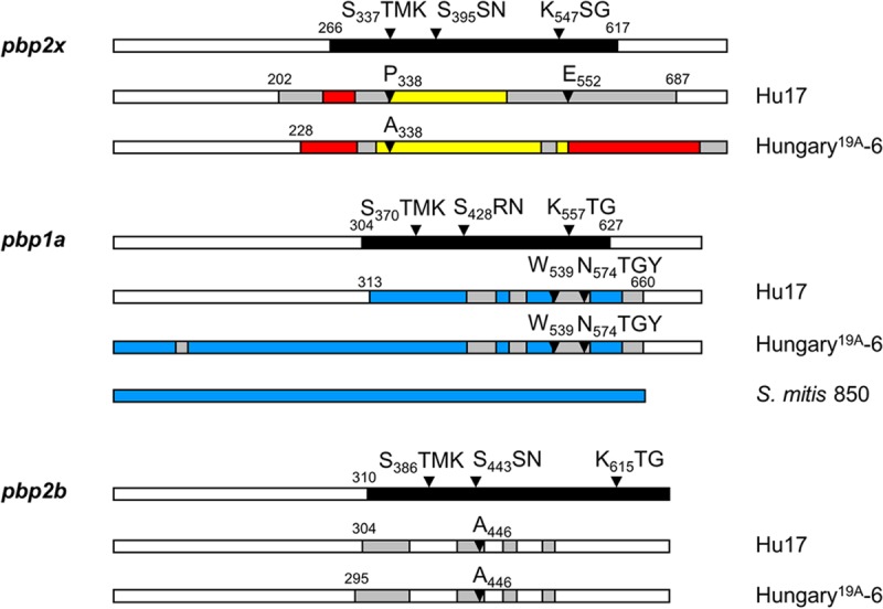
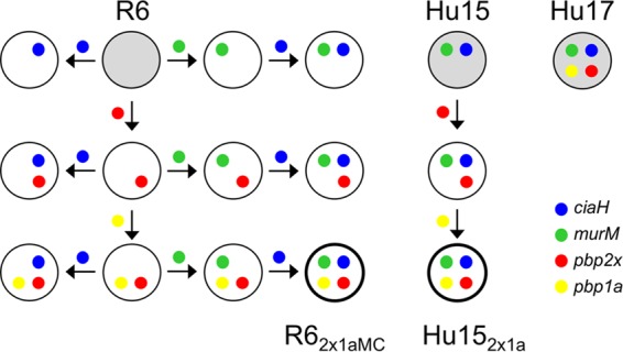
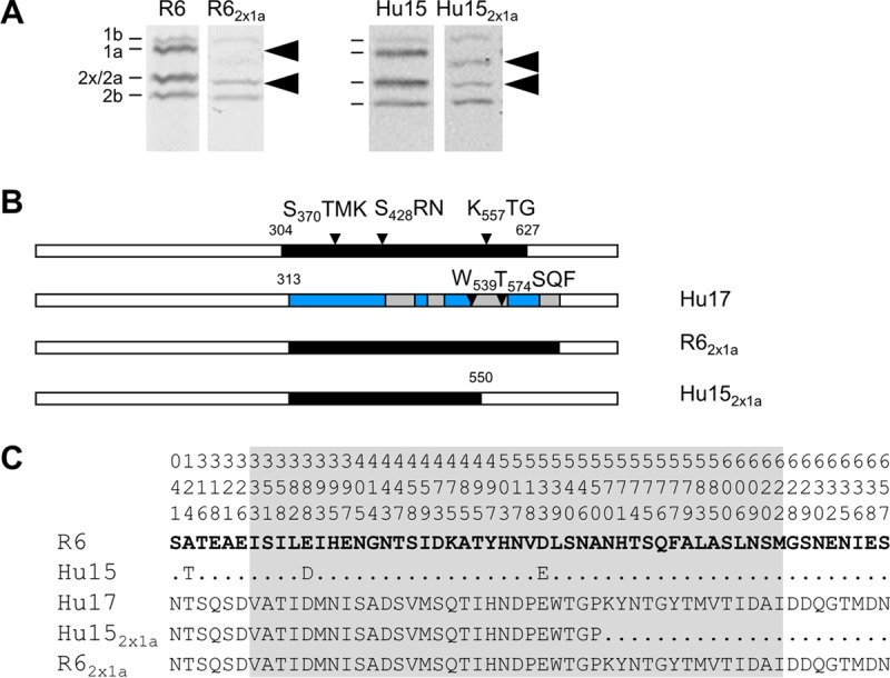
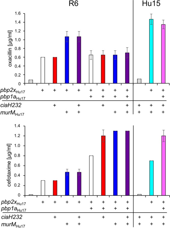
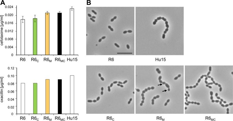
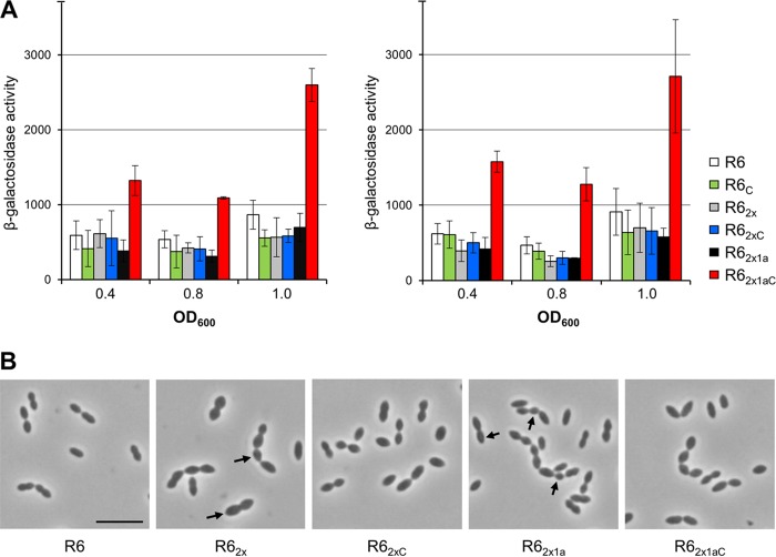
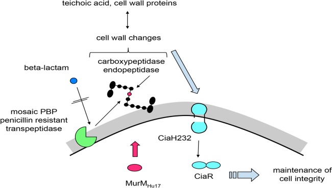
The Streptococcus pneumoniae clone Hungary19A-6 expresses unusually high levels of β-lactam resistance, which is in part due to mutations in the MurM gene, encoding a transferase involved in the synthesis of branched peptidoglycan. Moreover, it contains the allele ciaH232, encoding the histidine kinase CiaH (M. Müller, P. Marx, R. Hakenbeck, and R. Brückner, Microbiology 157:3104-3112, 2011, https://doi.org/10.1099/mic.0.053157-0). High-level penicillin resistance primarily requires the presence of low-affinity (mosaic) penicillin binding protein (PBP) genes, as, for example, in strain Hu17, a closely related member of the Hungary19A-6 lineage. Interestingly, strain Hu15 is β-lactam sensitive due to the absence of mosaic PBPs. This unique situation prompted us to investigate the development of cefotaxime resistance in transformation experiments with genes known to play a role in this phenotype, pbp2x, pbp1a, murM, and ciaH, and penicillin-sensitive recipient strains R6 and Hu15. Characterization of phenotypes, peptidoglycan composition, and CiaR-mediated gene expression revealed several novel aspects of penicillin resistance. The murM gene of strain Hu17 (murMHu17), which is highly similar to murM of Streptococcus mitis, induced morphological changes which were partly reversed by ciaH232. murMHu17 conferred cefotaxime resistance only in the presence of the pbp2x of strain Hu17 (pbp2xHu17). The ciaH232 allele contributed to a remarkable increase in cefotaxime resistance in combination with pbp2xHu17 and pbp1a of strain Hu17 (pbp1aHu17), accompanied by higher levels of expression of CiaR-regulated genes, documenting that ciaH232 responds to PBP1aHu17-mediated changes in cell wall synthesis. Most importantly, the proportion of branched peptides relative to the proportion of linear muropeptides increased in cells containing mosaic PBPs, suggesting an altered enzymatic activity of these proteins.

Keywords: CiaH; MurM; PBP1a; PBP2x; Streptococcus pneumoniae; peptidoglycan analysis.


Figures

FIG 1
FIG 1
Mosaic PBPs of S. pneumoniae Hu17 and Hungary19A-6. Mosaic gene structures were deduced by comparison of the reference PBP2x sequences of strains S. pneumoniae R6 (white) and S. mitis M3 (red), NCTC10712 (yellow), and 850 (blue). Highly similar sequences (<5% difference) are shown in the same color; gray areas are divergent sequences of unknown origin. The numbers indicate the codons defining the sequence blocks that diverge from the pbp2xR6 nucleotide sequence by >15%. The domain structure and active-site boxes are indicated on top; the black area represents the transpeptidase domain. Mutations potentially relevant for the resistance phenotype are indicated.
FIG 2
FIG 2
Schematic representation of transformation steps. The genes of PBP2x, PBP1a, MurM, and CiaH present in penicillin-resistant strain Hu17 (top right) were introduced individually or in various combinations into sensitive nonencapsulated laboratory strain S. pneumoniae R6 or into clinical isolate Hu15, as indicated by the colors. The direction of gene transfer is indicated by arrows. Gray circles represent recipient strains R6 and Hu15 and donor strain Hu17. Transformants containing all four genes are identified at the bottom and named according to the order of the transformation steps.
FIG 3
FIG 3
PBP2x variants obtained in transformation experiments. (A) PBP profiles of transformants containing mosaic PBP2x. PBPs were visualized after incubation with BocillinFL followed by separation by SDS-PAGE and fluorography. Recipient strains S. pneumoniae R6 and Hu15 were included for comparison, as indicated on top. The positions of the PBPs are marked on the left side. Arrowheads, low-affinity PBP2x. (B) Schematic representation of pbp2x in transformants R62x and Hu152x compared to strains R6 (white sequence blocks) and Hu17 (black sequence blocks). (Top row) The transpeptidase domain is shown in black, and the active-site motifs are indicated. (C) Deduced peptide sequences of PBP2x of the transformants compared to the peptide sequences of PBP2x of the penicillin-sensitive strain S. pneumoniae R6 and the donor PBP2x of Hu17. The first three lines indicate the amino acid positions. Only the residues at positions with altered residues are shown; residues identical to those in strain R6 (bold letters) are indicated by dots. The transpeptidase domain is shaded gray.
FIG 4
FIG 4
PBP1a variants obtained in transformation experiments. (A) PBP profiles of transformants containing mosaic PBP1a. PBPs were visualized after incubation with BocillinFL followed by separation by SDS-PAGE and fluorography. Recipient strains S. pneumoniae R6 and Hu15 were included for comparison, as indicated on top. The positions of the PBPs are marked on the left side. Arrowheads, low-affinity PBP2x and PBP1a. (B) Schematic representation of pbp1a in transformants compared to strains R6 (white sequence blocks) and Hu17 (black sequence blocks). (Top row) The transpeptidase domain is shown in black, and the active-site motifs are indicated. (C) Deduced peptide sequences of PBP1a of the transformants compared to the peptide sequences of PBP1a of the penicillin-sensitive strain S. pneumoniae R6 and the donor PBP1a of Hu17. The first three lines indicate the amino acid positions. Only the residues at positions with altered residues are shown; residues identical to those in strain R6 (bold letters) are indicated by dots. The transpeptidase domain is shaded gray.
FIG 5
FIG 5
Resistance patterns of various S. pneumoniae transformants. The two β-lactams oxacillin and cefotaxime were used. Mean values from at least three independent experiments are shown. Bars indicate standard deviations. The MIC values of oxacillin and cefotaxime for the donor strain Hu17 were 30 μg/ml and 1.6 μg/ml, respectively.
FIG 6
FIG 6
Cell morphology and resistance pattern of S. pneumoniae R6 transformants carrying the murM and/or ciaH gene from strain Hu17. (A) The MIC values for the transformants are indicated as colored bars, and those for control strains R6 and Hu15 are indicated as white bars. The two β-lactams cefotaxime and oxacillin were used. Mean values from at least three independent experiments are shown. Bars indicate standard deviations. (B) Representative phase-contrast micrographs are shown. The strains were grown in C+Y medium. Arrows, altered cell morphology. Bar, 5 μm.
FIG 7
FIG 7
β-Galactosidase activities expressed from CiaR-regulated promoters and cell morphology of S. pneumoniae R6 derivatives carrying the ciaH232 allele. (A) Strains were grown in C+Y medium. β-Galactosidase activities were determined at three different time points and are given in nanomoles of nitrophenol produced per minute and milligram of protein. Mean values and standard deviations from three independent experiments are presented. The activities of the CiaR-dependent promoters in the R62x1aC strain were significantly different (P < 0.05) from those of the promoters in the other strains. The promoters and relevant genetic markers are indicated. (B) Phase-contrast images taken at the mid-exponential growth phase. The strains were grown in C+Y medium. Arrows, altered cell morphology. Bar, 5 μm.
FIG 8
FIG 8
Schematic view of the interactions between MurM, PBPs, and the CiaRH system. Black, a branched muropeptide; red, l-Ala in the interpeptide bridge added by MurMHu17; gray: cell wall; black line, cell membrane. See the text for details.

References

    1. Högberg L, Geli P, Ringberg H, Melander E, Lipsitch M, Ekdahl K. 2007. Age- and serogroup-related differences in observed durations of nasopharyngeal carriage of penicillin-resistant pneumococci. J Clin Microbiol 45:948–952. doi: 10.1128/JCM.01913-06. - DOI - PMC - PubMed
    1. Short KR, Diavatopoulos DA. 2015. Nasopharyngeal colonization with Streptococcus pneumoniae, p 279–291. In Brown J, Hammerschmidt S, Orihuela CJ (ed), Streptococcus pneumoniae molecular mechanisms of host-pathogen interactions. Academic Press, London, United Kingdom.
    1. O'Brien KL, Wolfson LJ, Watt JP, Henkle E, Deloria-Knoll M, McCall N, Lee E, Mulholland K, Levine OS, Cherian T, Hib and Pneumococcal Global Burden of Disease Study Team. 2009. Burden of disease caused by Streptococcus pneumoniae in children younger than 5 years: global estimates. Lancet 374:893–902. doi: 10.1016/S0140-6736(09)61204-6. - DOI - PubMed
    1. Henriques-Normark B, Tuomanen E. 2013. The pneumococcus: epidemiology, microbiology, and pathogenesis. Cold Spring Harb Perspect Med 3:a010215. doi: 10.1101/cshperspect.a010215. - DOI - PMC - PubMed
    1. McGee L, McDougal L, Zhou J, Spratt BG, Tenover FC, George R, Hakenbeck R, Hryniewicz W, Lefévre JC, Tomasz A, Klugman KP. 2001. Nomenclature of major antimicrobial-resistant clones of Streptococcus pneumoniae defined by the pneumococcal molecular epidemiology network. J Clin Microbiol 39:2565–2571. doi: 10.1128/JCM.39.7.2565-2571.2001. - DOI - PMC - PubMed

Publication types

MeSH terms

LinkOut - more resources