. 2017 Jun 7;37(23):5620-5633.
doi: 10.1523/JNEUROSCI.2617-16.2017. Epub 2017 May 8.

Uncoupling of UNC5C with Polymerized TUBB3 in Microtubules Mediates Netrin-1 Repulsion

Affiliations

Uncoupling of UNC5C with Polymerized TUBB3 in Microtubules Mediates Netrin-1 Repulsion

Qiangqiang Shao et al. J Neurosci. .

Abstract

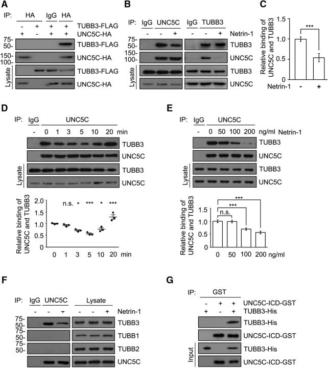
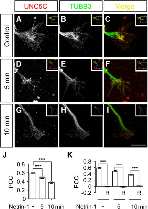
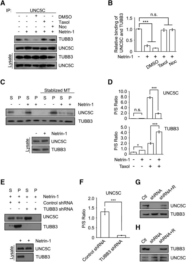
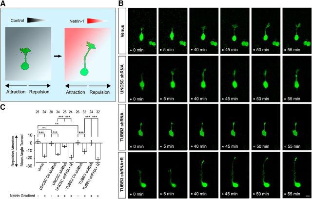
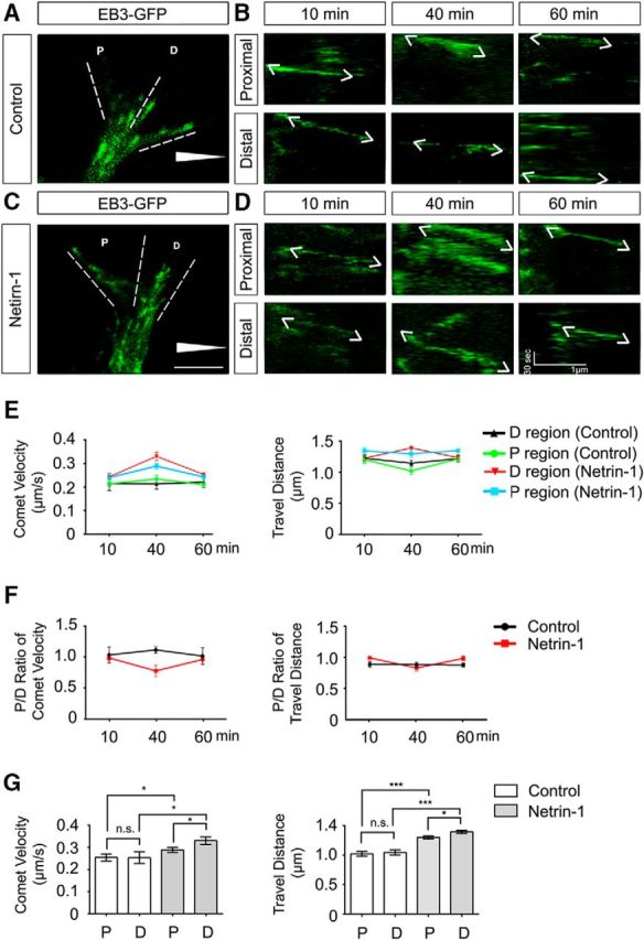
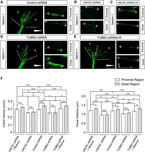
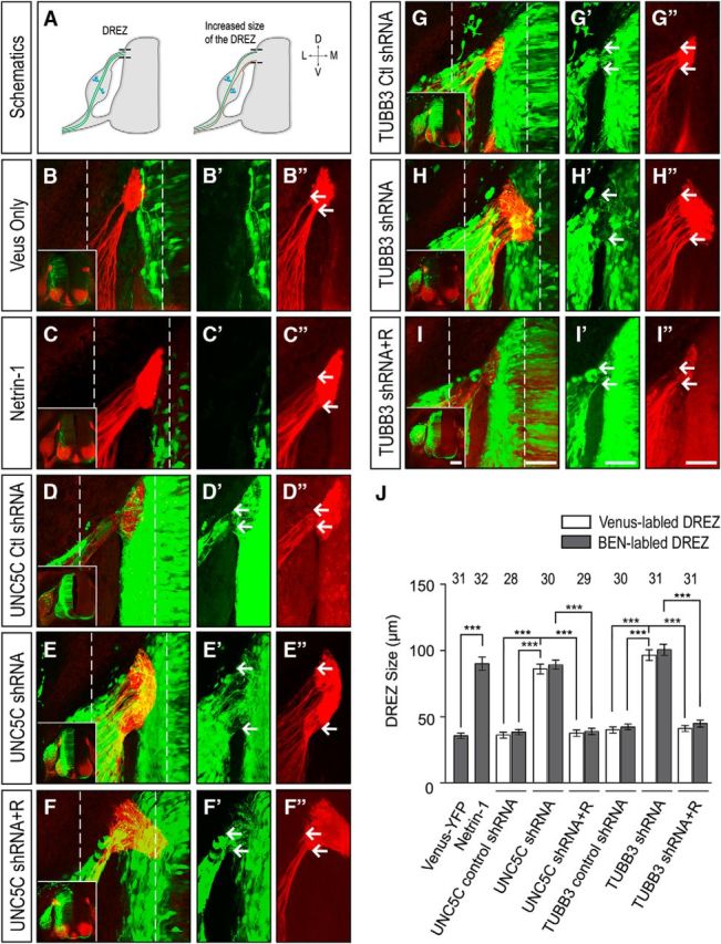
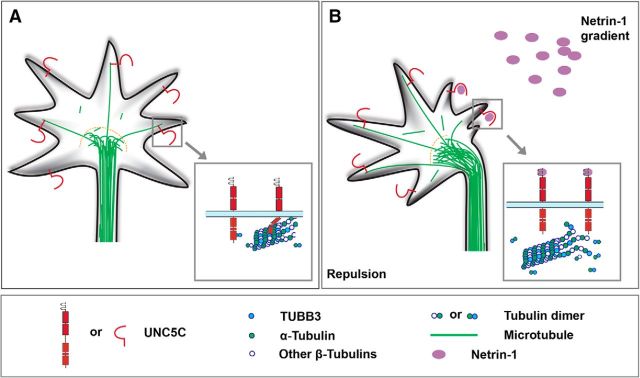
Modulation of microtubule (MT) dynamics is a key event of cytoskeleton remodeling in the growth cone (GC) during axon outgrowth and pathfinding. Our previous studies have shown that the direct interaction of netrin receptor DCC and DSCAM with polymerized TUBB3, a neuron-specific MT subunit in the brain, is required for netrin-1-mediated axon outgrowth, branching, and attraction. Here, we show that uncoupling of polymerized TUBB3 with netrin-1-repulsive receptor UNC5C is involved in netrin-1-mediated axonal repulsion. TUBB3 directly interacted with UNC5C and partially colocalized with UNC5C in the peripheral area of the GC of primary neurons from the cerebellar external granule layer of P2 mouse pups of both sexes. Netrin-1 reduced this interaction as well as the colocalization of UNC5C and TUBB3 in the GC. Results from the in vitro cosedimentation assay indicated that UNC5C interacted with polymerized TUBB3 in MTs and netrin-1 decreased this interaction. Knockdown of either TUBB3 or UNC5C blocked netrin-1-promoted axon repulsion in vitro and caused defects in axon projection of DRG toward the spinal cord in vivo Furthermore, live-cell imaging of end-binding protein 3 tagged with EGFP (EB3-GFP) in primary external granule layer cells showed that netrin-1 differentially increased MT dynamics in the GC with more MT growth in the distal than the proximal region of the GC during repulsion, and knockdown of either UNC5C or TUBB3 abolished the netrin-1 effect. Together, these data indicate that the disengagement of UNC5C with polymerized TUBB3 plays an essential role in netrin-1/UNC5C-mediated axon repulsion.SIGNIFICANCE STATEMENT Proper regulation of microtubule (MT) dynamics in the growth cone plays an important role in axon guidance. However, whether guidance cues modulate MT dynamics directly or indirectly is unclear. Here, we report that dissociation of UNC5C and polymerized TUBB3, the highly dynamic β-tubulin isoform in neurons, is essential for netrin-1/UNC5C-promoted axon repulsion. These results not only provide a working model of direct modulation of MTs by guidance cues in growth cone navigation but also help us to understand molecular mechanisms underlying developmental brain disorders associated with TUBB3 mutations.

Keywords: TUBB3; UNC5C; axon guidance; growth cone; microtubule dynamics; netrin.


Figures

Figure 1.
Figure 1.
Netrin-1 reduces the interaction of UNC5C with TUBB3. A, UNC5C interacts with TUBB3 in HeLa cells. TUBB3-FLAG was cotransfected with UNC5C-HA into HeLa cells. Anti-HA (UNC5C) precipitated TUBB3-FLAG. B, Netrin-1 decreases the interaction of endogenous TUBB3 with UNC5C. Dissociated neurons from P4 mouse cerebella were stimulated with either the control or netrin-1-conditioned media (netrin-1 concentration was ∼300 ng/μl) for 5 min. Cell lysates were immunoprecipitated with either anti-UNC5C (left) or with anti-TUBB3 (right), and the immunoblot was analyzed with anti-TUBB3 and anti-UNC5C. C, Quantification of B from three independent experiments. ***p < 0.001 (two-tailed Student's t test). D, E, Netrin-1 regulates the interaction of endogenous UNC5C with TUBB3 in a time-dependent (D) and dose-dependent manner (E). Primary neurons from P4 mouse cerebella were stimulated with either netrin-1-conditioned media (netrin-1 concentration was ∼300 ng/μl) for 0–20 min (D) or purified netrin-1 for 5 min (E). Bottom, Quantification. *p < 0.05 (one-way ANOVA and Tukey's test for post hoc comparisons). ***p < 0.001 (one-way ANOVA and Tukey's test for post hoc comparisons). F, Endogenous UNC5C interacts with TUBB3, not TUBB1 and TUBB2, in response of netrin-1 treatment. Primary neurons from P4 mouse cerebella were stimulated with either the control or netrin-1-conditioned media (netrin-1 concentration was ∼300 ng/μl) for 5 min. Cell lysates were immunoprecipitated with anti-UNC5C and followed by probing with anti-TUBB1, anti-TUBB2, or anti-TUBB3. G, Direct interaction of TUBB3 with UNC5C. Purified TUBB3 was incubated with purified intracellular domain of UNC5C tagged with GST in vitro. The anti-GST antibody was used to immunoprecipitate proteins, and the blot was analyzed with anti-TUBB3.
Figure 2.
Figure 2.
Netrin-1 reduces the subcellular overlap of UNC5C with TUBB3 in the GC of primary neurons. A–C, Overlap of immunofluorescent signals of UNC5C (A) and TUBB3 (B) in the GC of mouse EGL cells. P2 mouse EGL neurons were cultured for 20 h and stimulated with a sham-purified control. C, Merged image of A, B. A–C, The value of PCC is 0.59 ± 0.02. D–I, Overlap of endogenous UNC5C with TUBB3 in the GC of EGL neurons was reduced after netrin-1 stimulation for either 5 min (D–F) or 10 min (G–I). F, I, Merged images of D, E and G, H, respectively. D–F and G–I, The value of PCC of UNC5C and TUBB3 is 0.49 ± 0.02 and 0.37 ± 0.02, respectively. Scale bar, 10 μm. J, Quantitative analysis of PCC in the control and netrin-1 groups. A total of 37 GCs in each group were analyzed. ***p < 0.001 (one-way ANOVA and Tukey's test for post hoc comparisons). K, Quantification of PCC after 90 degree counterclockwise rotation of one fluorescence channel. ***p < 0.001 (one-way ANOVA and Tukey's test for post hoc comparisons). R, Rotation.
Figure 3.
Figure 3.
Netrin-1 decreases the interaction of UNC5C with polymerized TUBB3. A, Taxol and nocodazole (Noc) inhibited the netrin-1-induced UNC5C/TUBB3 dissociation. Dissociated P4 mouse cerebellar neurons were treated with purified netrin-1 in the presence of 1 μm Taxol, 3 μm nocodazole, or DMSO. B, Quantification of A from three independent experiments showing relative binding of UNC5C to TUBB3. ***p < 0.001 (one-way ANOVA and Tukey's test for post hoc comparisons). C, Primary P4 mouse cerebellar neurons were stimulated with netrin-1 or sham-purified control, and a cosedimentation assay of cell lysates was performed in the absence or presence of Taxol. UNC5C and TUBB3 in the pellet (P) and supernatant (S) fractions were examined by immunoblotting using anti-UNC5C and anti-TUBB3 antibodies, respectively. D, Quantification of C from three independent experiments showing P/S ratio of UNC5C and TUBB3. *p < 0.05 (one-way ANOVA and Tukey's test for post hoc comparisons). ***p < 0.001 (one-way ANOVA and Tukey's test for post hoc comparisons). E, Dissociated P4 mouse cerebellar neurons were transfected with either TUBB3 shRNA or control shRNA and stimulated with purified netrin-1. The cosedimentation assay was conducted with Taxol to stabilize MTs in vitro as above. F, Quantification of P/S ratio of E from three independent experiments. ***p < 0.001 (two-tailed Student's t test). G, Knockdown of endogenous UNC5C in primary neurons. P4 mouse cerebellar neurons were nucleofected with control shRNA, UNC5C shRNA, or UNC5C shRNA plus wild-type human UNC5C. Ctl, Control shRNA; R, wild-type human UNC5C. H, TUBB3 shRNA specifically knocked down endogenous TUBB3. P4 mouse cerebellar neurons were transfected with control shRNA, TUBB3 shRNA, or TUBB3 plus wild-type human TUBB3. Ctl, Control shRNA; R, wild-type human TUBB3.
Figure 4.
Figure 4.
TUBB3 is required for netrin-1/UNC5C-mediated axon repulsion. A, Schematic diagram of axon turning of P2 mouse EGL cells in a control (left) or netrin-1 (right) gradient using a Dunn chamber axon guidance assay. B, Live-cell imaging showing axon turning of P2 EGL cells transfected with Venus-YFP only or combination of Venus-YFP with other indicated constructs. Nucleofection of Venus-YFP into P2 EGL neurons allowed visualization of axon projection. A left-to-right netrin-1 gradient was established as shown in A. Scale bar, 10 μm. C, Quantification of axon turning of P2 EGL neurons. Data are mean ± SEM. ***p < 0.001 (one-way ANOVA and Tukey's test for post hoc comparisons). The numbers on the top of each bar indicate the numbers of GCs analyzed in the corresponding groups.
Figure 5.
Figure 5.
Visualization of MT dynamics in the GC via time-lapse recording of EB3-GFP in primary neurons during axon turning. A–D, Trajectories of EB3-GFP comets in the GC of P2 mouse EGL neurons. P2 mouse EGL neurons were dissociated and nucleofected with EB3-GFP. Primary neurons were cultured for 2 d, and a Dunn chamber axon guidance assay was performed in the absence (A, B) or presence of a netrin-1 gradient (C, D). B, D, Kymographs of EB3-GFP comet movements in the proximal (upper) and distal regions (lower) of the GCs in A and C, respectively. Scale bar, 10 μm. White triangle represents either a control or netrin-1 gradient. E, F, Quantification of velocity (E, left) and travel distance (E, right) as well as ratios of velocity (F, left) and travel distance (F, right) of moving EB3-GFP comets in proximal and distal regions of the GC of P2 EGL cells (A–D). G, Bar graph comparisons of the velocity (left) and travel distance (right) of moving EB3-GFP comets in the GC of P2 EGL cells at 40 min after image capture from A–D. Data are mean ± SEM (3 GCs in each group). *p < 0.05 (one-way ANOVA with Tukey's test for post hoc comparisons). ***p < 0.001 (one-way ANOVA with Tukey's test for post hoc comparisons).
Figure 6.
Figure 6.
TUBB3 is specifically involved in netrin-1/UNC5C-regulated MT dynamics in the GC of P2 cerebellar EGL neurons. A–E, P2 mouse EGL neurons were cotransfected EB3-GFP with control shRNA (A), UNC5C shRNA (B), UNC5C shRNA plus wild-type human UNC5C (C), TUBB3 shRNA (D), or TUBB3 shRNA plus wild-type human TUBB3 (E), respectively, and a Dunn chamber axon guidance assay was performed. A, D, E, Left, Live-cell imaging tracking EB3-GFP comet movements in the GC. Right, Kymographs of EB3-GFP comets in the proximal (upper) and distal (lower) regions of the GC. B, C, Kymographs of EB3-GFP comet movements in the proximal (upper) and distal (lower) regions of the GC of P2 cerebellar EGL cells transfected with UNC5C shRNA (B) or UNC5C shRNA plus wild-type human UNC5C (C). Scale bar, 10 μm. White triangle represents either a control or netrin-1 gradient. F, Quantification of the velocity (left) and travel distance (right) of moving EB3-GFP comets in the GC of P2 EGL cells (A–E) at 40 min after image capture. Data are mean ± SEM (3 GCs in each group). *p < 0.05 (one-way ANOVA with Tukey's test for post hoc comparisons). ***p < 0.001 (one-way ANOVA with Tukey's test for post hoc comparisons).
Figure 7.
Figure 7.
TUBB3 is essential for DRG axon projection in vivo. A, Schematic showing DRG axon projection toward the chick spinal cord after electroporation. Brown represents DRG axon bundles with projection deficiency. Green represents normal DRG axon bundles. B–C″, The chick neural tube at Stage 16 was electroporated with Venus YFP only (B–B″) or Venus YFP plus AP-netrin-1 (C–C″), and transverse sections of the spinal cord were stained with BEN antibody (red). D–I″, The chick neural tube at Stages 12–15 was electroporated with Venus YFP plus UNC5C control shRNA (D–D″), Venus YFP plus UNC5C shRNA (E–E″), Venus YFP plus UNC5C shRNA and wild-type human UNC5C (F–F″), Venus YFP plus TUBB3 control shRNA (G–G″), Venus YFP plus TUBB3 shRNA (H–H″), Venus YFP plus TUBB3 shRNA and wild-type human TUBB3 (I–I″). Transverse sections of the Venus YFP-labeled spinal cords were stained with BEN antibody. B–I, Overlay confocal images of green (Venus YFP) and red (BEN staining) fluorescence. Insets, Corresponding low-magnification images of B–I. B′–I′, B″–I″, Venus YFP images and images of BEN antibody immunostaining of the region of interest in B–I (dashed lines), respectively. Scale bar, 50 μm. J, Quantification of DREZ size. ***p < 0.001 (one-way ANOVA with Tukey's test for post hoc comparisons). Both Venus YFP and BEN antibody staining consistently demarcates DRG axon projections with no statistically significant difference in DREZ size in each group. The numbers on the top of each bar indicate the numbers of samples tested in the corresponding groups. R, Rescue constructs.
Figure 8.
Figure 8.
A working model of direct involvement of MT dynamics in netrin-1-mediated axon repulsion. A, A typical GC of developing neurons with lamelliopodia and finger-like filopodia. In the peripheral region of the GC, UNC5C interacts with TUBB3 in “pioneer polymerized MTs” in the absence of netrin-1 gradient. B, Binding of netrin-1 to UNC5C on the side of GC close to the netrin-1 gradient differentially reduces the interaction of UNC5C with polymerized TUBB3 in MTs and triggers GC collapse on that side resulting in axon repulsion, despite the fact that netrin-1 induces MT polyermization/dynamics in the GC.

Similar articles

Cited by

References

    1. Ackerman SL, Kozak LP, Przyborski SA, Rund LA, Boyer BB, Knowles BB (1997) The mouse rostral cerebellar malformation gene encodes an UNC-5-like protein. Nature 386:838–842. 10.1038/386838a0 - DOI - PubMed
    1. Alcántara S, Ruiz M, De Castro F, Soriano E, Sotelo C (2000) Netrin 1 acts as an attractive or as a repulsive cue for distinct migrating neurons during the development of the cerebellar system. Development 127:1359–1372. - PubMed
    1. Avraham O, Hadas Y, Vald L, Hong S, Song MR, Klar A (2010) Motor and dorsal root ganglion axons serve as choice points for the ipsilateral turning of dI3 axons. J Neurosci 30:15546–15557. 10.1523/JNEUROSCI.2380-10.2010 - DOI - PMC - PubMed
    1. Bartoe JL, McKenna WL, Quan TK, Stafford BK, Moore JA, Xia J, Takamiya K, Huganir RL, Hinck L (2006) Protein interacting with C-kinase 1/protein kinase Calpha-mediated endocytosis converts netrin-1-mediated repulsion to attraction. J Neurosci 26:3192–3205. 10.1523/JNEUROSCI.3469-05.2006 - DOI - PMC - PubMed
    1. Bronner-Fraser M, Fraser S (1989) Developmental potential of avian trunk neural crest cells in situ. Neuron 3:755–766. 10.1016/0896-6273(89)90244-4 - DOI - PubMed

Publication types