. 2017 May 23;114(21):E4241-E4250.
doi: 10.1073/pnas.1621511114. Epub 2017 May 8.

Hypoxia treatment reverses neurodegenerative disease in a mouse model of Leigh syndrome

Affiliations

Hypoxia treatment reverses neurodegenerative disease in a mouse model of Leigh syndrome

Michele Ferrari et al. Proc Natl Acad Sci U S A. .

Erratum in

Abstract

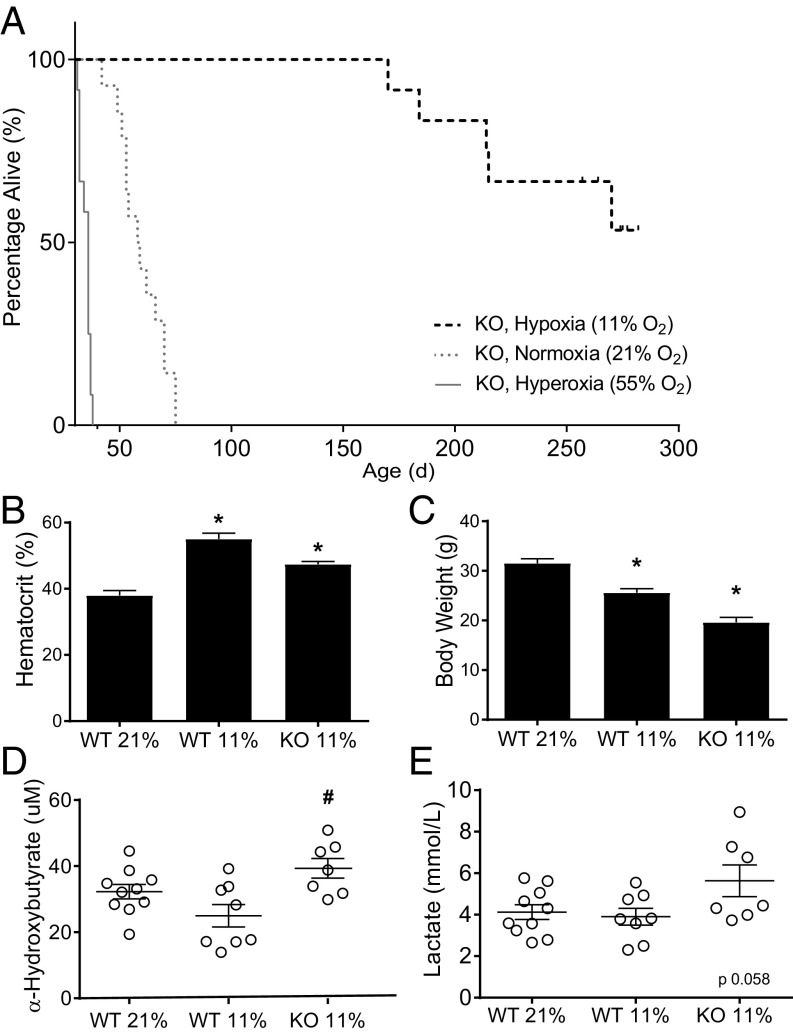
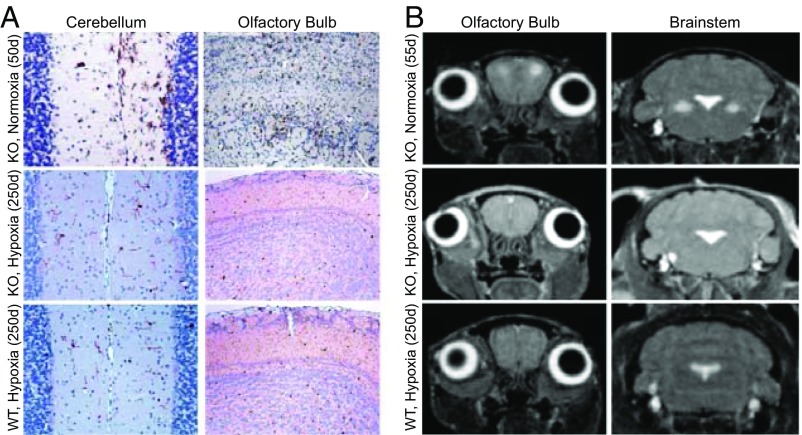
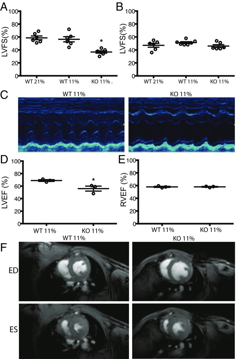
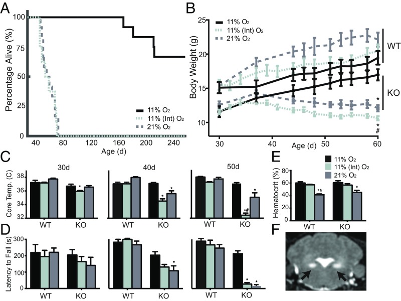
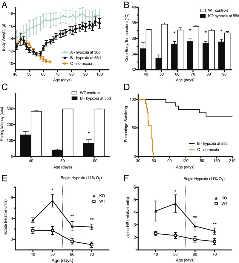
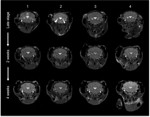
The most common pediatric mitochondrial disease is Leigh syndrome, an episodic, subacute neurodegeneration that can lead to death within the first few years of life, for which there are no proven general therapies. Mice lacking the complex I subunit, Ndufs4, develop a fatal progressive encephalopathy resembling Leigh syndrome and die at ≈60 d of age. We previously reported that continuously breathing normobaric 11% O2 from an early age prevents neurological disease and dramatically improves survival in these mice. Here, we report three advances. First, we report updated survival curves and organ pathology in Ndufs4 KO mice exposed to hypoxia or hyperoxia. Whereas normoxia-treated KO mice die from neurodegeneration at about 60 d, hypoxia-treated mice eventually die at about 270 d, likely from cardiac disease, and hyperoxia-treated mice die within days from acute pulmonary edema. Second, we report that more conservative hypoxia regimens, such as continuous normobaric 17% O2 or intermittent hypoxia, are ineffective in preventing neuropathology. Finally, we show that breathing normobaric 11% O2 in mice with late-stage encephalopathy reverses their established neurological disease, evidenced by improved behavior, circulating disease biomarkers, and survival rates. Importantly, the pathognomonic MRI brain lesions and neurohistopathologic findings are reversed after 4 wk of hypoxia. Upon return to normoxia, Ndufs4 KO mice die within days. Future work is required to determine if hypoxia can be used to prevent and reverse neurodegeneration in other animal models, and to determine if it can be provided in a safe and practical manner to allow in-hospital human therapeutic trials.

Keywords: Leigh syndrome; hypoxia; mitochondria; neurodegeneration; oxygen.


Conflict of interest statement

Conflict of interest statement: V.K.M., W.M.Z., and I.H.J. are listed as coinventors on a patent application submitted by Massachusetts General Hospital on the therapeutic uses of hypoxia for mitochondrial disease.

Figures

Fig. 1.
Fig. 1.
Survival, hematocrit, and disease markers in older Ndufs4 KO mice breathing 11% O2. (A) Survival rates of mice breathing various oxygen levels. Hypoxia-treated Ndufs4 KO mice were all alive at 170 d (n = 12). The experiment was interrupted once 50% survival was achieved. All WT mice breathing 11% and 21% O2 (n = 14 per group) survived for 300 d (Kaplan–Meier log-rank between KO at 11% O2 and KO at 21% O2, P < 0.0001, HR, 7.58; 95% CI, 2.75–20.9, n = 12 per group). (B) After 30 wk of treatment, hematocrit was elevated in KO (n = 7) and WT mice (n = 8) breathing 11% O2. (C) Body weight of KO (n = 7) and WT (n = 9) controls at age 250 d. (D and E) Plasma levels of α-hydroxybutyrate (D) and lactate (E) in hypoxic KO mice at age 250 d, compared with age-matched WT mice breathing 21% or 11% O2. Data are mean ± SD. *P < 0.05 vs. WT, 21% O2; #P < 0.05 vs. WT, 11% O2, one-way ANOVA with Bonferroni’s correction.
Fig. 2.
Fig. 2.
Absence of neurodegenerative pathology in 250-d-old hypoxia-treated Ndufs4 KO mice. (A) Representative images with staining for the microglial activation marker Iba-1 (n = 3). Normoxic KO mice at 50 d show a significant inflammatory response in the cerebellum and OB. Analogous images in 250-d-old hypoxic KO mice and WT mice do not show brain inflammation. (B) Axial head MRI images showing bilateral, symmetrical hyperintense lesions in the OB (Left) and brainstem (Right) of normoxic-breathing KO mice at 55 d. These lesions were not present in hypoxic KO mice at 250 d.
Fig. 3.
Fig. 3.
Cardiac ultrasound (US) and cardiac MRI reveal depressed LV myocardial function in Ndufs4 KO mice breathing 11% O2 at age >200 d. (A) US of LV fractional shortening (LVFS) of mice breathing various oxygen concentrations at age 200 d (n = 6). (B) US LVFS of mice breathing various oxygen concentrations at 50 d of age (n = 6). (C) Representative M-mode scans of the left ventricle in WT and KO mice breathing 11% O2 at 200 d. (D) MRI showing LV ejection fraction of WT and KO mice breathing 11% O2 at 200 d of age (n = 3). (E) 9.4-T MRI showing right ventricular ejection fraction in WT and KO mice breathing 11% O2 at 200 d of age (n = 3). (F) Representative MRI sections of WT and KO mice breathing 11% O2 at 200 d of age. Images display end-diastolic (ED) and end-systolic (ES) reconstructions. Scans were obtained during sedation with isoflurane while breathing 21% O2. Data are mean ± SD. *P < 0.05 vs. WT, one-way ANOVA with Bonferroni's correction for multiple comparisons.
Fig. 4.
Fig. 4.
Detrimental effects of breathing 55% O2 on lungs and OB of Ndufs4 KO mice. (A) Pulmonary WD ratio of KO and WT mice breathing various oxygen levels. Lungs were weighed after 24 h of breathing 55% O2 at age 30 d (n = 7). (B) Myeloperoxidase activity in the lungs of mice after 24 h of breathing 55% O2 at age 30 d (n = 7). (C) H&E staining of lungs exposed to 55% O2 for 48 h at age 30 d (n = 3, representative images; see text for description). (D) Axial MRI scans of murine brains showing the OB (Upper) and brainstem (BS; Lower) of KO mice breathing 21% O2 at 60 d and Ndufs4 KO mice breathing 55% O2 for 24 h at 30 d (representative images; n = 3). Data are mean ± SD. *P < 0.05 vs. other groups, one-way ANOVA with Bonferroni's correction.
Fig. 5.
Fig. 5.
Intermittent hypoxic (11%) breathing (10 h/d) does not alleviate mitochondrial disease in Ndufs4 KO mice. (A) Survival rates for Ndufs4 KO mice with intermittent hypoxic breathing (Int) vs. those breathing 21% O2 (log-rank P = 0.77; HR, 1.13; 95% CI, 0.47–2.73; n = 8). (B) Body weights after breathing at various oxygen levels and during intermittent hypoxic breathing starting at age 30 d (n = 8). (C and D) Core temperature (C) and falling latency (D) from an accelerating, rotating rod for KO mice breathing various oxygen levels or subjected to intermittent hypoxic breathing starting at 30 d of age (n = 8). (E) Hematocrit levels in WT and KO mice following 3 wk of exposure to normoxic, hypoxic, or intermittent hypoxic breathing (n = 4). (F) Representative MRI in a 60-d-old KO mouse exposed to intermittent hypoxic breathing. Arrows denote lesions in vestibular nuclei. Data are mean ± SD. *P < 0.05 vs. KO breathing 11% O2; #P < 0.05 vs. KO breathing 21% O2.
Fig. 6.
Fig. 6.
Breathing at moderate hypoxia (17% O2) does not alleviate mitochondrial disease. (A) Survival rates for Ndufs4 mice breathing various oxygen levels starting at age 30 d. (B and C) Time course of body weight (B) and body temperature (C) for mice breathing 17% O2 and those breathing breathing 11% or 21% O2 for 30, 40, and 50 d (n = 6). (D) Venous hematocrit values after 3 wk of exposure to various oxygen levels (n = 6). Data are mean ± SD. *P < 0.05 vs. 11% oxygen, #P < 0.05 vs. 17% oxygen.
Fig. 7.
Fig. 7.
Hypoxic breathing (11% O2) rescues survival, body weight, and behavior of Ndufs4 mice with late-stage neurologic impairment. (A) Growth curves of Ndufs4 KO mice exposed to early hypoxic breathing (group A) starting at 30 d of age (gray triangles), those exposed to late hypoxic breathing (group B) starting at 55 d of age (black circles), and control Ndufs4 KO mice with normoxic breathing (group C; orange squares) (B and C) Body temperature (B) and latency of falling from an accelerating rotating rod (C) in Ndufs4 KO mice with late-stage disease (group B) and WT controls exposed to breathing 11% O2 starting at 55 d of age (n = 8). (D) Survival rates of hypoxic breathing mice with late-stage disease treated with hypoxia at age 55 d (group B; black, n = 17) and controls breathing air (group C; orange, n = 13) (log-rank P < 0.0001). Data are mean ± SD. *P < 0.05 vs. 50 d. (E and F) Lactate (E) and α-hydroxybutyrate (F) time course in group B mice and WT controls. *P < 0.05, **P > 0.05 compared with WT at 40 d.
Fig. 8.
Fig. 8.
Breathing 11% O2 in late-stage neurological disease reverses the radiographic lesions of Ndufs4 mice seen on MRI. Four Ndufs4 mice were breathing 21% O2 until they developed late-stage neurological disease (55 d). They underwent MRI to document bilateral lesions in the vestibular nuclei (Upper, white arrow). Subsequently they commenced breathing 11% oxygen. The mice were scanned again at 2 wk and 4 wk of hypoxic breathing (Middle and Lower, respectively). Neurologic lesions disappeared by 4 wk of hypoxic breathing. The section of the fourth ventricle shown at the center of the brainstem appeared enlarged in the early scans, suggesting parenchymal atrophy. After 4 wk of treatment, this area appeared to be reduced in size, and morphological relationships were restored.
Fig. 9.
Fig. 9.
Breathing 11% O2 in late-stage neurological disease reverses pathological inflammation in the brains of Ndufs4 KO mice. Representative images with Iba-1 staining of the OB and cerebellum (CB) in KO mice and WT controls (n = 3 per group). Iba-1 is a marker of inflammation in the brain, indicative of microglial activation. Images demonstrate the reversibility of the neuropathological pattern by breathing 11% O2 at the late stage of disease (55 d). (A) KO mice breathing 21% O2. (B) KO mice breathing 21% O2 up to 55 d and then breathing 11% O2 (to 160 d). MRI-demonstrated reversal of the lesions reported in Fig. 8 was observed in these same mice. (C) Normoxic WT controls. (D) Hypoxic WT controls.

References

    1. Vafai SB, Mootha VK. Mitochondrial disorders as windows into an ancient organelle. Nature. 2012;491:374–383. - PubMed
    1. Gorman GS, et al. Prevalence of nuclear and mitochondrial DNA mutations related to adult mitochondrial disease. Ann Neurol. 2015;77:753–759. - PMC - PubMed
    1. Alston CL, Rocha MC, Lax NZ, Turnbull DM, Taylor RW. The genetics and pathology of mitochondrial disease. J Pathol. 2017;241:236–250. - PMC - PubMed
    1. Lake NJ, Compton AG, Rahman S, Thorburn DR. Leigh syndrome: One disorder, more than 75 monogenic causes. Ann Neurol. 2016;79:190–203. - PubMed
    1. Debs R, et al. Biotin-responsive basal ganglia disease in ethnic Europeans with novel SLC19A3 mutations. Arch Neurol. 2010;67:126–130. - PubMed

Publication types