

A molecular beacon-based approach for live-cell imaging of RNA transcripts with minimal target engineering at the single-molecule level
- PMID: 28484218
- PMCID: PMC5431543
- DOI: 10.1038/s41598-017-01740-1
A molecular beacon-based approach for live-cell imaging of RNA transcripts with minimal target engineering at the single-molecule level
Abstract
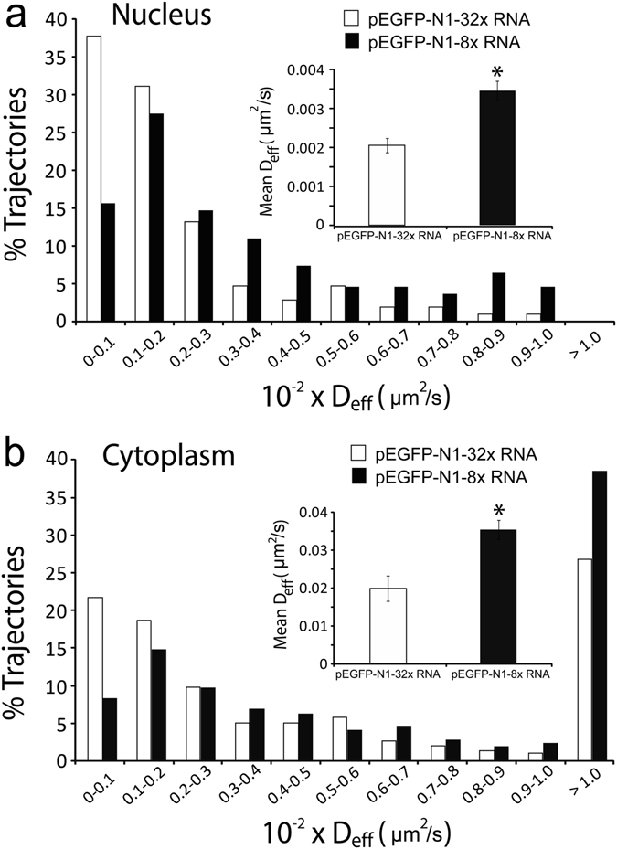
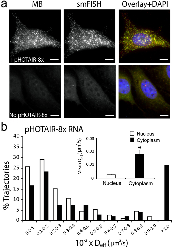
Analysis of RNA dynamics and localization at the single-molecule level in living cells has been predominantly achieved by engineering target RNAs with large insertions of tandem repeat sequences that are bound by protein-based or oligonucleotide-based fluorescent probes. Thus, individual RNAs are tagged by multiple fluorescent probes, making them detectable by fluorescence microscopy. Since large insertions may affect RNA processes including trafficking and localization, here we present a strategy to visualize single RNA transcripts in living cells using molecular beacons (MBs) - fluorogenic oligonucleotide probes - with minimal target engineering. The MBs are composed of 2'-O-methyl RNAs with a fully phosphorothioate-modified loop domain (2Me/PSLOOP MBs), an architecture that elicits marginal levels of nonspecific signals in cells. We showed that MBs can detect single transcripts containing as few as 8 target repeat sequences with ~90% accuracy. In both the nucleus and the cytoplasm, mRNAs harboring 8 repeats moved faster than those with 32 repeats, suggesting that intracellular activities are less impeded by smaller engineered insertions. We then report the first MB-based imaging of intracellular dynamics and localization of single long noncoding RNAs (lncRNAs). We envision the proposed minimally-engineered, MB-based technology for live-cell single-molecule RNA imaging could facilitate new discoveries in RNA research.
Conflict of interest statement
The authors declare that they have no competing interests.
Figures


Similar articles
-
Single-molecule detection and tracking of RNA transcripts in living cells using phosphorothioate-optimized 2'-O-methyl RNA molecular beacons.Biomaterials. 2016 Sep;100:172-83. doi: 10.1016/j.biomaterials.2016.05.022. Epub 2016 May 24. Biomaterials. 2016. PMID: 27261815
-
Optimizing Molecular Beacons for Intracellular Analysis of RNA.Methods Mol Biol. 2018;1649:243-257. doi: 10.1007/978-1-4939-7213-5_16. Methods Mol Biol. 2018. PMID: 29130202
-
Live-Cell Imaging of Long Noncoding RNAs Using Molecular Beacons.Methods Mol Biol. 2019;2038:21-33. doi: 10.1007/978-1-4939-9674-2_2. Methods Mol Biol. 2019. PMID: 31407275
-
Engineering Novel Molecular Beacon Constructs to Study Intracellular RNA Dynamics and Localization.Genomics Proteomics Bioinformatics. 2017 Oct;15(5):279-286. doi: 10.1016/j.gpb.2017.04.004. Epub 2017 Sep 21. Genomics Proteomics Bioinformatics. 2017. PMID: 28942262 Free PMC article. Review.
-
Nanostructured probes for RNA detection in living cells.Ann Biomed Eng. 2006 Jan;34(1):39-50. doi: 10.1007/s10439-005-9003-6. Epub 2006 Feb 7. Ann Biomed Eng. 2006. PMID: 16463087 Review.
Cited by
-
Electrophoretic mobility shift as a molecular beacon-based readout for miRNA detection.Biosens Bioelectron. 2021 Oct 1;189:113307. doi: 10.1016/j.bios.2021.113307. Epub 2021 May 15. Biosens Bioelectron. 2021. PMID: 34062334 Free PMC article.
-
Imaging of DNA and RNA in Living Eukaryotic Cells to Reveal Spatiotemporal Dynamics of Gene Expression.Annu Rev Biochem. 2020 Jun 20;89:159-187. doi: 10.1146/annurev-biochem-011520-104955. Epub 2020 Mar 16. Annu Rev Biochem. 2020. PMID: 32176523 Free PMC article. Review.
-
Single-molecule analysis of endogenous β-actin mRNA trafficking reveals a mechanism for compartmentalized mRNA localization in axons.Proc Natl Acad Sci U S A. 2018 Oct 9;115(41):E9697-E9706. doi: 10.1073/pnas.1806189115. Epub 2018 Sep 25. Proc Natl Acad Sci U S A. 2018. PMID: 30254174 Free PMC article.
-
Subcellular Spatial Transcriptomes: Emerging Frontier for Understanding Gene Regulation.Cold Spring Harb Symp Quant Biol. 2019;84:31-45. doi: 10.1101/sqb.2019.84.040352. Epub 2020 Jun 1. Cold Spring Harb Symp Quant Biol. 2019. PMID: 32482897 Free PMC article.
-
Time-lapse imaging of microRNA activity reveals the kinetics of microRNA activation in single living cells.Sci Rep. 2017 Oct 3;7(1):12642. doi: 10.1038/s41598-017-12879-2. Sci Rep. 2017. PMID: 28974737 Free PMC article.
References
Publication types
MeSH terms
Substances
LinkOut - more resources
Full Text Sources
Other Literature Sources
Research Materials
Miscellaneous
