. 2017 May 8;7(1):1575.
doi: 10.1038/s41598-017-01805-1.

Glial cell-line derived neurotrophic factor protects human islets from nutrient deprivation and endoplasmic reticulum stress induced apoptosis

Affiliations

Glial cell-line derived neurotrophic factor protects human islets from nutrient deprivation and endoplasmic reticulum stress induced apoptosis

Shadab Abadpour et al. Sci Rep. .

Abstract

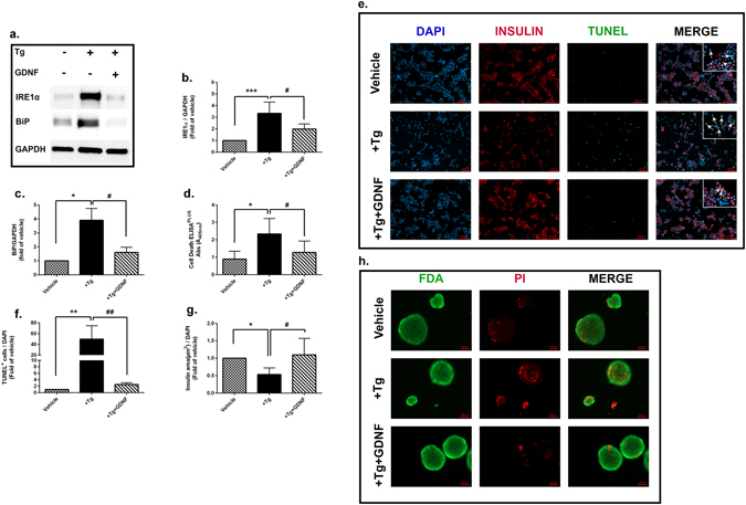
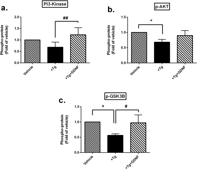
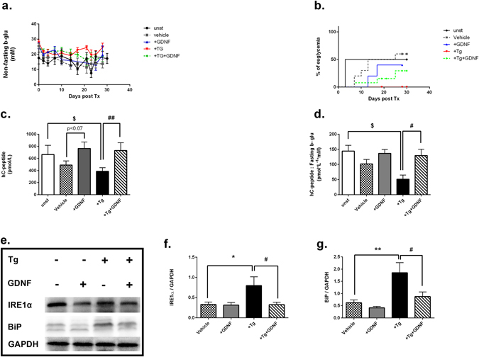
One of the key limitations to successful human islet transplantation is loss of islets due to stress responses pre- and post-transplantation. Nutrient deprivation and ER stress have been identified as important mechanisms leading to apoptosis. Glial Cell-line Derived Neurotrophic Factor (GDNF) has recently been found to promote islet survival after isolation. However, whether GDNF could rescue human islets from nutrient deprivation and ER stress-mediated apoptosis is unknown. Herein, by mimicking those conditions in vitro, we have shown that GDNF significantly improved glucose stimulated insulin secretion, reduced apoptosis and proinsulin:insulin ratio in nutrient deprived human islets. Furthermore, GDNF alleviated thapsigargin-induced ER stress evidenced by reduced expressions of IRE1α and BiP and consequently apoptosis. Importantly, this was associated with an increase in phosphorylation of PI3K/AKT and GSK3B signaling pathway. Transplantation of ER stressed human islets pre-treated with GDNF under kidney capsule of diabetic mice resulted in reduced expressions of IRE1α and BiP in human islet grafts with improved grafts function shown by higher levels of human C-peptide post-transplantation. We suggest that GDNF has protective and anti-apoptotic effects on nutrient deprived and ER stress activated human islets and could play a significant role in rescuing human islets from stress responses.


Conflict of interest statement

S.O.G is employed by AstraZeneca.

Figures

Figure 1
Figure 1
GDNF improves human islets function and viability under nutrient deprived culture condition. (a) Insulin secretion in response to basal (1.67 mM) and stimulated (20 mM) level of glucose (GSIS) (b) calculated as stimulation index in human islets prior to experiment start (0 hrs, unst islets) and after treatment for 72 hrs with or without GDNF (200 ng/nl) under nutrient deprived culture condition, n = 6. (c) Secreted insulin and proinsulin in culture medium were measured by EIA and presented as the proinsulin to insulin ratio, n = 4. (d) Apoptosis evaluated by cell death ELISAPLUS in human islets, n = 6. (e) Representative images showing insulin (red) and TUNEL (green) with DAPI nuclear staining (blue) of dispersed human islets treated with or without GDNF. (f) Ratio of insulin area as well as (g) score of TUNEL+ cells over nuclear staining. Data is presented as a fold of vehicle islets, n = 3, Five images were taken from every slide and minimum of 2000 cells were scored. (h) Representative images showing intact islets stained for PI (red) and FDA (green) prior to islets culture (0 hrs) and after 72 hrs culture of nutrient deprived islets treated with or without GDNF. Statistical analysis: p-values were analyzed by Wilcoxon matched-pairs test in (a), nonparametric ANOVA with Dunn’s corrections in (b) (d), Mann-Whitney U-test in (c,f,g). For all analysis, data is presented as mean ± SD. *p < 0.05 vs.unst islets, #0.05, ##p < 0.01, ###p < 0.001 vs. vehicle islets, *p < 0.05 vs stimulated (20 mM) glucose. Unst: unstarved islets. The n refers to the number of independent donors used for each experiment.
Figure 2
Figure 2
GDNF reduces ER stress and consequently ER stress induced apoptosis in human islets. (a) Representative western blot of IRE1α and BiP in human islets treated with Tg (1 µm) with or without GDNF (200 ng/ml) for 48 hrs. (b,c) Quantification of IRE1α and BiP band densities normalized to housekeeping gene GAPDH. Data is presented as a fold of vehicle islets, n = 6. (d) Cell death analysis by cell death ELISAPLUS of human islets treated with Tg with or without GDNF for 48 hrs, n = 9. (e) Representative images showing insulin (red), TUNEL (green) and DAPI (blue) nuclear staining of dispersed human islets treated with Tg with or without GDNF for 48 hrs. (f) Score of TUNEL+ cells (g) and measurement of insulin area to DAPI nuclear staining in dispersed human islets. Data is presented as a fold of vehicle islets. n = 3, five images were taken from each slide and minimum of 2000 cells were scored. (h) Representative images showing intact islets stained for PI (red) and FDA (green). For all analysis, data is presented as mean ± SD and p-values were analyzed by nonparametric ANOVA with Dunn’s corrections.*p < 0.05, **p < 0.01, ***p < 0.001 vs. vehicle islets, #p < 0.05, ##p < 0.01 vs. Tg-treated islets. Tg: Thapsigargin. The n refers to number of independent donors used for each experiment.
Figure 3
Figure 3
GDNF protective effect on ER stress induced human islets is through activation of PI3K/AKT signaling pathway. (a–c) Assessment of phosphoproteins in the PI3K/AKT/GSK3B signaling pathway by multiplex assay in human islets treated with Tg (1 µm) with or without GDNF (200 ng/ml) for 48 hrs. Data is presented as a fold of vehicle islets. n = 6. For all analysis, data is presented as mean ± SD and p-values were analyzed by nonparametric ANOVA with Dunn’s corrections. *p < 0.05vs. vehicle islets, #p < 0.05, ##p < 0.01 vs. Tg-treated islets. Tg:Thapsigargin. The n refers to number of donors used for each experiment.
Figure 4
Figure 4
GDNF improves islet grafts function and reduces ER stress post transplantation. (a) Random blood glucose levels in diabetic mice transplanted with human islets pre-cultured for 48 hrs in complete medium (unst, n = 6), nutrient deprived medium (vehicle, n = 11) with GDNF (200 ng/ml, n = 6), Tg (1 µM, n = 8), or Tg+GDNF(n = 8). (b) Percentage of euglycemic mice post transplantation. (c) Circulating levels of hC-peptide measured by EIA in plasma at day 30 post transplantation (d) and presented as ratio of hC-peptide over fasting b-glu. (e) Representative western blot for protein expression of IRE1α and BiP in human islet grafts harvested on day 30 post transplantation. (f,g) Quantification of IRE1α and BiP bands densities normalized to housekeeping gene GAPDH. For all analysis, data is presented as mean ± SD and p-values were analyzed by nonparametric ANOVA with Dunn’s corrections or Mann-Whitney U-test. The n refers to number of mice in each experimental group, *P < 0.05, **p < 0.01 vs. vehicle islets, #p < 0.05, ##p < 0.01 vs. Tg-treated islets. $p < 0.05 vs unstv group. Log-rank (Mantel-Cox) test was used to analyze the difference in percentage of euglycemic animals post transplantation. Unst: unstarved, Tg: Thapsigargin, B-glu: Blood glucose, hCpeptide: human C-peptide.

Similar articles

Cited by

References

    1. Cooke DW, Plotnick L. Type 1 diabetes mellitus in pediatrics. Pediatrics in review/American Academy of Pediatrics. 2008;29:374–384. doi: 10.1542/pir.29-11-374. - DOI - PubMed
    1. Kincaid MM, Cooper AA. Misfolded proteins traffic from the endoplasmic reticulum (ER) due to ER export signals. Molecular biology of the cell. 2007;18:455–463. doi: 10.1091/mbc.E06-08-0696. - DOI - PMC - PubMed
    1. Wang J, et al. A mutation in the insulin 2 gene induces diabetes with severe pancreatic beta-cell dysfunction in the Mody mouse. The Journal of clinical investigation. 1999;103:27–37. doi: 10.1172/JCI4431. - DOI - PMC - PubMed
    1. Butler AE, et al. Beta-cell deficit and increased beta-cell apoptosis in humans with type 2 diabetes. Diabetes. 2003;52:102–110. doi: 10.2337/diabetes.52.1.102. - DOI - PubMed
    1. Eizirik DL, Cardozo AK, Cnop M. The role for endoplasmic reticulum stress in diabetes mellitus. Endocrine reviews. 2008;29:42–61. doi: 10.1210/er.2007-0015. - DOI - PubMed

Publication types

MeSH terms

Substances

LinkOut - more resources