

Differential Targeting of Hsp70 Heat Shock Proteins HSPA6 and HSPA1A with Components of a Protein Disaggregation/Refolding Machine in Differentiated Human Neuronal Cells following Thermal Stress
- PMID: 28484369
- PMCID: PMC5401876
- DOI: 10.3389/fnins.2017.00227
Differential Targeting of Hsp70 Heat Shock Proteins HSPA6 and HSPA1A with Components of a Protein Disaggregation/Refolding Machine in Differentiated Human Neuronal Cells following Thermal Stress
Abstract
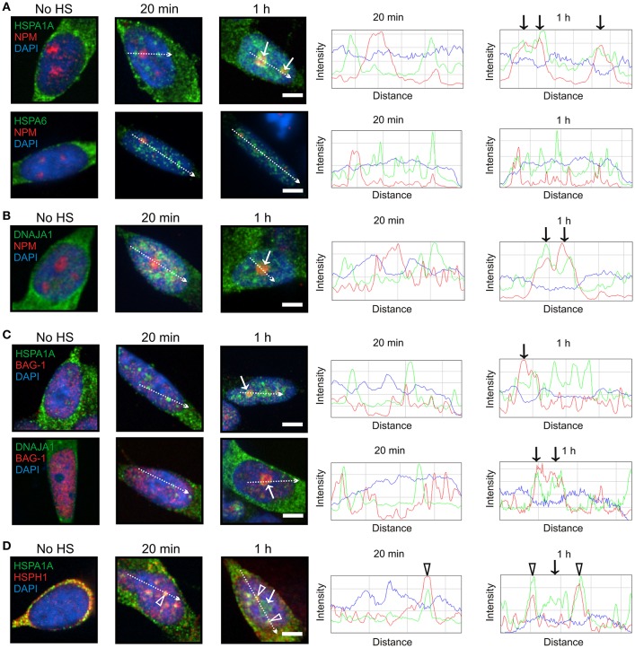
Heat shock proteins (Hsps) co-operate in multi-protein machines that counter protein misfolding and aggregation and involve DNAJ (Hsp40), HSPA (Hsp70), and HSPH (Hsp105α). The HSPA family is a multigene family composed of inducible and constitutively expressed members. Inducible HSPA6 (Hsp70B') is found in the human genome but not in the genomes of mouse and rat. To advance knowledge of this little studied HSPA member, the targeting of HSPA6 to stress-sensitive neuronal sites with components of a disaggregation/refolding machine was investigated following thermal stress. HSPA6 targeted the periphery of nuclear speckles (perispeckles) that have been characterized as sites of transcription. However, HSPA6 did not co-localize at perispeckles with DNAJB1 (Hsp40-1) or HSPH1 (Hsp105α). At 3 h after heat shock, HSPA6 co-localized with these members of the disaggregation/refolding machine at the granular component (GC) of the nucleolus. Inducible HSPA1A (Hsp70-1) and constitutively expressed HSPA8 (Hsc70) co-localized at nuclear speckles with components of the machine immediately after heat shock, and at the GC layer of the nucleolus at 1 h with DNAJA1 and BAG-1. These results suggest that HSPA6 exhibits targeting features that are not apparent for HSPA1A and HSPA8.
Keywords: DNAJ (Hsp40); HSPA1A (Hsp70-1); HSPA6 (Hsp70B'); HSPA8 (Hsc70); HSPH1 (Hsp105α); human neuronal SH-SY5Y cells.
Figures


Similar articles
-
Components of a mammalian protein disaggregation/refolding machine are targeted to nuclear speckles following thermal stress in differentiated human neuronal cells.Cell Stress Chaperones. 2017 Mar;22(2):191-200. doi: 10.1007/s12192-016-0753-x. Epub 2016 Dec 13. Cell Stress Chaperones. 2017. PMID: 27966060 Free PMC article.
-
Localization of heat shock protein HSPA6 (HSP70B') to sites of transcription in cultured differentiated human neuronal cells following thermal stress.J Neurochem. 2014 Dec;131(6):743-54. doi: 10.1111/jnc.12970. Epub 2014 Dec 2. J Neurochem. 2014. PMID: 25319762
-
Intracellular Targeting of Heat Shock Proteins in Differentiated Human Neuronal Cells Following Proteotoxic Stress.J Alzheimers Dis. 2018;66(3):1295-1308. doi: 10.3233/JAD-180536. J Alzheimers Dis. 2018. PMID: 30412487
-
Structural and functional diversities between members of the human HSPB, HSPH, HSPA, and DNAJ chaperone families.Biochemistry. 2008 Jul 8;47(27):7001-11. doi: 10.1021/bi800639z. Epub 2008 Jun 17. Biochemistry. 2008. PMID: 18557634 Review.
-
HSPA6 and its role in cancers and other diseases.Mol Biol Rep. 2022 Nov;49(11):10565-10577. doi: 10.1007/s11033-022-07641-5. Epub 2022 Jun 6. Mol Biol Rep. 2022. PMID: 35666422 Review.
Cited by
-
Endogenous retroviruses are dysregulated in ALS.iScience. 2024 May 28;27(7):110147. doi: 10.1016/j.isci.2024.110147. eCollection 2024 Jul 19. iScience. 2024. PMID: 38989463 Free PMC article.
-
HSPA2 Chaperone Contributes to the Maintenance of Epithelial Phenotype of Human Bronchial Epithelial Cells but Has Non-Essential Role in Supporting Malignant Features of Non-Small Cell Lung Carcinoma, MCF7, and HeLa Cancer Cells.Cancers (Basel). 2020 Sep 24;12(10):2749. doi: 10.3390/cancers12102749. Cancers (Basel). 2020. PMID: 32987811 Free PMC article.
-
Similar Normalizing Effect of HSP70 and YB-1 Stress Proteins on the Brain Transcription of a Mouse Model of Alzheimer's Disease.Mol Neurobiol. 2025 Jun 24. doi: 10.1007/s12035-025-05135-6. Online ahead of print. Mol Neurobiol. 2025. PMID: 40553235
-
Alterations in Proteostasis System Components in Peripheral Blood Mononuclear Cells in Parkinson Disease: Focusing on the HSP70 and p62 Levels.Biomolecules. 2022 Mar 24;12(4):493. doi: 10.3390/biom12040493. Biomolecules. 2022. PMID: 35454081 Free PMC article.
-
Tau Protein Squired by Molecular Chaperones During Alzheimer's Disease.J Mol Neurosci. 2018 Nov;66(3):356-368. doi: 10.1007/s12031-018-1174-3. Epub 2018 Sep 28. J Mol Neurosci. 2018. PMID: 30267382 Review.
References
-
- Asea A. A., Brown I. R. (2008). Heat Shock Proteins and the Brain: Implications for Neurodegenerative Diseases and Neuroprotection, eds Asea A. A., Brown I. R. (New York, NY: Springer Science+Business Media, B.V; ), 1–371.
LinkOut - more resources
Full Text Sources
Other Literature Sources
Miscellaneous