Review
. 2017 Apr 21:11:23.
doi: 10.3389/fnsys.2017.00023. eCollection 2017.

Activity-Dependent Synaptic Refinement: New Insights from Drosophila

Affiliations
Review

Activity-Dependent Synaptic Refinement: New Insights from Drosophila

Fernando Vonhoff et al. Front Syst Neurosci. .

Abstract

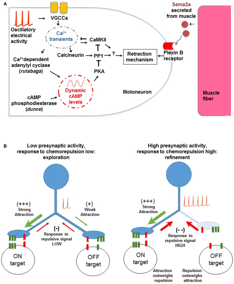
During development, neurons establish inappropriate connections as they seek out their synaptic partners, resulting in supernumerary synapses that must be pruned away. The removal of miswired synapses usually involves electrical activity, often through a Hebbian spike-timing mechanism. A novel form of activity-dependent refinement is used by Drosophila that may be non-Hebbian, and is critical for generating the precise connectivity observed in that system. In Drosophila, motoneurons use both glutamate and the biogenic amine octopamine for neurotransmission, and the muscle fibers receive multiple synaptic inputs. Motoneuron growth cones respond in a time-regulated fashion to multiple chemotropic signals arising from their postsynaptic partners. Central to this mechanism is a very low frequency (<0.03 Hz) oscillation of presynaptic cytoplasmic calcium, that regulates and coordinates the action of multiple downstream effectors involved in the withdrawal from off-target contacts. Low frequency calcium oscillations are widely observed in developing neural circuits in mammals, and have been shown to be critical for normal connectivity in a variety of neural systems. In Drosophila these mechanisms allow the growth cone to sample widely among possible synaptic partners, evaluate opponent chemotropic signals, and withdraw from off-target contacts. It is possible that the underlying molecular mechanisms are conserved widely among invertebrates and vertebrates.

Keywords: chemorepulsion; neuromuscular junction; non-Hebbian; oscillation; second messengers.


Figures

Figure 1
Figure 1
The events associated with synaptic targeting at the Drosophila NMJ. (A) Initial motoneuron projections make filopodial contacts (green) onto both the target muscle as well as to multiple off-target muscle fibers (Halpern et al., ; Sink and Whitington, 1991). (B) During normal development, off-target contacts are withdrawn, leading to the final specific connectivity (green). The refinement must occur during an early critical period and depends on presynaptic electrical activity (Jarecki and Keshishian, ; White et al., ; Carrillo et al., 2010). (C) When neural activity is suppressed, the off-target contacts are retained (red), leading to ectopic synapses. The transition from a growth cone filopodium to a synapse is rapid and the refinement of ectopic contacts occurs while growth cones are still motile, consistent with the critical period for refinement at the Drosophila NMJ. Ectopic contacts that fail to withdraw develop into functional synapses. This is in contrast to scenarios observed in other systems where synaptic contacts have to be stabilized and then refined by mechanisms that rely on prolonged period of synaptic competition.
Figure 2
Figure 2
The molecular and cellular mechanisms involved in synaptic refinement. (A) The interactions were identified by genetic tests and transgenic manipulations. A low frequency voltage oscillation activates voltage gated Ca2+ channels (VGCCs). The resulting Ca2+ entry regulates Ca2+-dependent effectors including Ca2+/calmodulin-dependent serine/threonine kinase II (CaMKII), Calcineurin (CaN), and Rutabaga. The latter increases cAMP levels, which in turn regulate PKA and PP1. The chemorepellant Sema2a is secreted by the muscle and activates the presynaptic PlexinB receptor. The response to Sema2a is gated by the level of presynaptic Ca2+ activity (see text for details). Arrows and T-shape lines indicate positive and negative regulation, respectively. The subcellular physical location and region of action of the molecular components have not been determined yet. (B) A model for non-Hebbian refinement at the Drosophila NMJ. The left panel shows an initial contact made by a motoneuron onto on-target and off-target muscle fibers. The molecular match is stronger with the on-target fiber. When Ca2+ levels are low, the response to the retrograde chemorepulsive signal from the muscle is muted, allowing the off-target contact to be retained. With neural activity and elevated presynaptic Ca2+ (right panel), the repulsive response is elevated, leading to the withdrawal of the off-target contact. Note that the model does not depend on correlated activity between the synaptic partners, as would be expected in a Hebbian mechanism.

Similar articles

Cited by

References

    1. Akerman C. J., Cline H. T. (2007). Refining the roles of GABAergic signaling during neural circuit formation. Trends Neurosci. 30, 382–389. 10.1016/j.tins.2007.06.002 - DOI - PubMed
    1. Amano M., Nakayama M., Kaibuchi K. (2010). Rho-kinase/ROCK: a key regulator of the cytoskeleton and cell polarity. Cytoskeleton 67, 545–554. 10.1002/cm.20472 - DOI - PMC - PubMed
    1. Ang L.-H., Chen W., Yao Y., Ozawa R., Tao E., Yonekura J., et al. . (2006). Lim kinase regulates the development of olfactory and neuromuscular synapses. Dev. Biol. 293, 178–190. 10.1016/j.ydbio.2006.01.030 - DOI - PubMed
    1. Arroyo D. A., Feller M. B. (2016). Spatiotemporal features of retinal waves instruct the wiring of the visual circuitry. Front. Neural Circuits 10:54. 10.3389/fncir.2016.00054 - DOI - PMC - PubMed
    1. Ayoob J. C., Terman J. R., Kolodkin A. L. (2006). Drosophila Plexin B is a Sema-2a receptor required for axon guidance. Development 133, 2125–2135. 10.1242/dev.02380 - DOI - PubMed

LinkOut - more resources