. 2017 Apr 25:9:111.
doi: 10.3389/fnagi.2017.00111. eCollection 2017.

Aging Impairs Hippocampal- Dependent Recognition Memory and LTP and Prevents the Associated RyR Up-regulation

Affiliations

Aging Impairs Hippocampal- Dependent Recognition Memory and LTP and Prevents the Associated RyR Up-regulation

Alejandra Arias-Cavieres et al. Front Aging Neurosci. .

Abstract

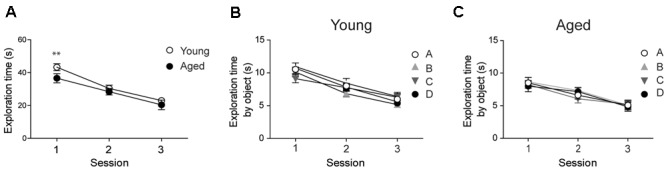
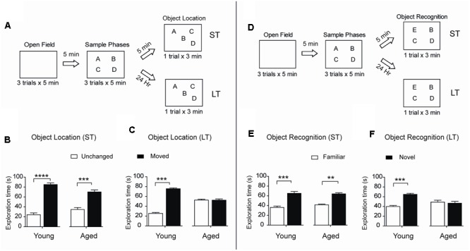
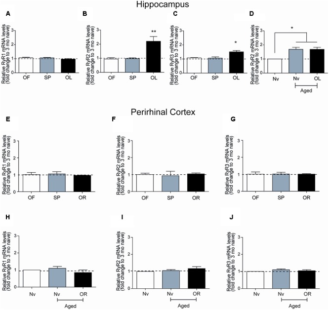
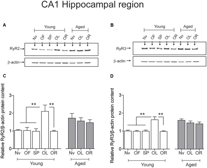
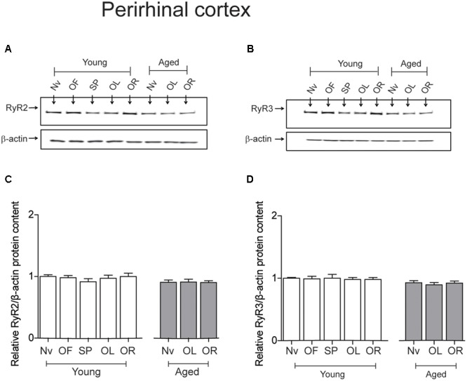
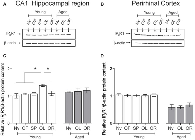
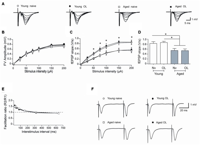
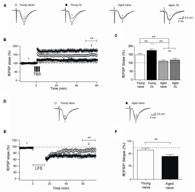
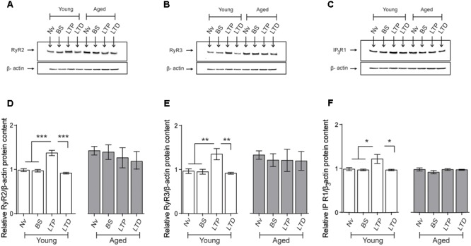
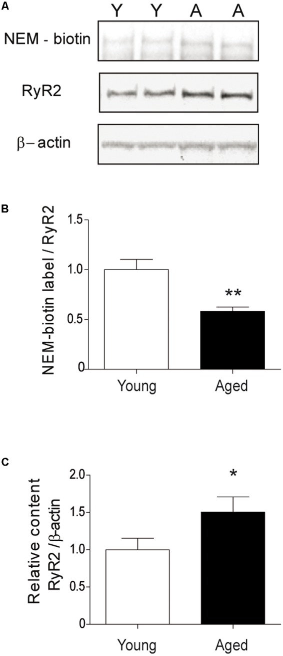
Recognition memory comprises recollection judgment and familiarity, two different processes that engage the hippocampus and the perirhinal cortex, respectively. Previous studies have shown that aged rodents display defective recognition memory and alterations in hippocampal synaptic plasticity. We report here that young rats efficiently performed at short-term (5 min) and long-term (24 h) hippocampus-associated object-location tasks and perirhinal cortex-related novel-object recognition tasks. In contrast, aged rats successfully performed the object-location and the novel-object recognition tasks only at short-term. In addition, aged rats displayed defective long-term potentiation (LTP) and enhanced long-term depression (LTD). Successful long-term performance of object-location but not of novel-object recognition tasks increased the protein levels of ryanodine receptor types-2/3 (RyR2/RyR3) and of IP3R1 in young rat hippocampus. Likewise, sustained LTP induction (1 h) significantly increased RyR2, RyR3 and IP3R1 protein levels in hippocampal slices from young rats. In contrast, LTD induction (1 h) did not modify the levels of these three proteins. Naïve (untrained) aged rats displayed higher RyR2/RyR3 hippocampal protein levels but similar IP3R1 protein content relative to young rats; these levels did not change following exposure to either memory recognition task or after LTP or LTD induction. The perirhinal cortex from young or aged rats did not display changes in the protein contents of RyR2, RyR3, and IP3R1 after exposure at long-term (24 h) to the object-location or the novel-object recognition tasks. Naïve aged rats displayed higher RyR2 channel oxidation levels in the hippocampus compared to naïve young rats. The RyR2/RyR3 up-regulation and the increased RyR2 oxidation levels exhibited by aged rat hippocampus are likely to generate anomalous calcium signals, which may contribute to the well-known impairments in hippocampal LTP and spatial memory that take place during aging.

Keywords: RyR oxidation; behavior; calcium release; gene expression; synaptic plasticity.


Figures

FIGURE 1
FIGURE 1
Young and aged rats explored the four objects for similar periods and did not exhibit preference for any given object: (A) At the second and third sessions, young (n = 44) and aged (n = 30) rats displayed similar average exploration times. Only at the first session the differences were statistically significant; ∗∗p < 0.01 [F(1,144)= 1.14]. (B) Young rats (n = 44) explored the four objects for equal times in each session. (C) Aged rats (n = 30) also explored the four objects for equal times in each session. In (B,C), differences within sessions were not statistically significant. Values represent Mean ± SEM. Statistical analysis was done with two-way ANOVA followed by Bonferroni’s post hoc test.
FIGURE 2
FIGURE 2
Aged rats display impairments in long-term spatial memory and novel-object recognition: (A) Diagram of the behavioral procedure used to test spatial memory retention in an object-location memory task. During the open field exploration phase, rats were habituated to the arena for 5 min during three consecutive days. In the sample phase, each rat was allowed to explore four different objects during 5 min for three consecutive sessions separated by 5 min intervals. Five minutes (ST: short-term assays) or 24 h (LT: long-term assays) after conclusion of the sample phase, independent groups of rats were exposed for 3 min to a new spatial arrangement, in which two of the four objects had different positions. (B) When tested after 5 min (ST), both young (n = 8), and aged (n = 7) rats discriminated the two spatially modified objects as evidenced by the increases in the time spent exploring the moved object. (C) When tested after 24 h (LT), young rats (n = 15) recognized the spatial rearrangement of objects and explored for longer times the moved object whereas aged rats (n = 8) did not. (D) Diagram of the behavioral procedures used to test novel-object recognition. The open field and the sample phases were as defined in (A); for further details, see Section “Materials and Methods.” Five min or 24 h after the conclusion of the sample phase, independent groups of rats were exposed for 3 min to a different object arrangement, in which a new object replaced one of the four original objects. (E) When tested after 5 min (ST), both young (n = 8), and aged (n = 7) rats explored for longer times the novel-object compared with the three familiar objects. (F) When tested after 24 h (LT), young rats (n = 13) discriminated and explored for longer times the novel-object while aged rats (n = 8) did not. Values represent Mean ± SEM. Statistical significance was assessed by the Student’s t-test (∗∗p < 0.01; ∗∗∗p < 0.005; ∗∗∗∗p < 0.001).
FIGURE 3
FIGURE 3
Ryanodine receptor mRNA levels in young or aged rats exposed to recognition tasks: qRT-PCR analysis of extracts from the CA1 hippocampal region or the PrhC isolated from young or aged rats was performed 6-h after long-term exposure to the object-location (OL) or the novel-object recognition (OR) tasks. All values for mRNA levels were normalized to the values displayed by naïve young rats. (A) CA1 RyR1 mRNA levels determined after the open field and the sample phases were similar to the values displayed by naïve young rats, and did not change after exposure to the OL task. (B) CA1 RyR2 mRNA levels determined after exposure of young rats to the open field and the sample phases were not statistically different to the values displayed by naïve young rats but increased after long-term exposure to the OL task. (C) CA1 RyR3 mRNA levels determined after exposure of young rats to the open field and the sample phases were similar to the values displayed by naïve young rats but increased after long-term exposure to the OL task. (D) In aged rats, RyR2 mRNA levels in the hippocampal CA1 region were higher than in naïve young rats and did not change after the OL task. In PrhC extracts from young or aged rats, RyR1 (E,H), RyR2 (F,I) and RyR3 (G,J) mRNA levels determined after the open field and the sample phases were similar to the values displayed by naïve young rats; these values did not change after exposure to the OR task. OF, open field; SP, sample phases. Values represent Mean ± SEM; n = 3 for each group. Statistical significance was assessed by One-way ANOVA followed by Tukey’s post hoc test (p < 0.05; ∗∗p < 0.01).
FIGURE 4
FIGURE 4
Effects of performing the recognition tasks on hippocampal RyR2/RyR3 protein contents: Representative immunoblots of RyR2 (A) and RyR3 (B) protein contents of extracts from the CA1 hippocampal region of young or aged rats; extracts were collected 6 h after long-term (24 h) exposure to the object-location (OL) or the novel-object recognition (OR) tasks. (C) In young rats, RyR2 protein content increased significantly after the OL [∗∗p < 0.01 (n = 7)] but not after the OR task (p > 0.05), and did not change in aged rats (n = 7) after exposure to either task (p > 0.05). (D) In young rats (n = 6), RyR3 protein content increased significantly in the CA1 region after the OL (∗∗p < 0.01) but not after the OR task (p > 0.05). Aged rats (n = 6) did not display changes in RyR3 protein content after performing either task (p > 0.05). OF, open field; SP, sample phases. Values represent Mean ± SEM. Statistical analysis was performed with one-way ANOVA followed by Tukey’s post hoc test.
FIGURE 5
FIGURE 5
Exposure to long-term memory tasks did not modify RyR2 and RyR3 protein contents in the PrhC from young or aged rats. (A,B) Representative immunoblots of RyR2 and RyR3 protein contents in PrhC extracts from young or aged rats. Extracts were collected from naïve rats, or from rats exposed at long-term to the object-location (OL) or the novel-object recognition (OR) tasks. (C) Naïve (Nv) young (n = 7) or aged rats (n = 7) and trained young (n = 7) or aged rats (n = 7) displayed similar RyR2 protein contents. (D) Naïve (Nv) young (n = 6) or aged rats (n = 6) and trained young (n = 6) or aged rats (n = 6) displayed similar RyR3 protein contents. OF, open field; SP, sample phases. Values represent Mean ± SEM. Statistical analysis was performed with one-way ANOVA followed by Tukey’s post hoc test.
FIGURE 6
FIGURE 6
Effects of performing the object location/novel-object recognition tasks on the IP3R1 protein content of the CA1 hippocampal region or the PrhC from young or aged rats: Representative immunoblots of the IP3R1 protein contents determined in (A), the CA1 hippocampal region or (B), the PrhC isolated from young or aged rats. All samples were collected 6 h after exposure at long-term to the object-location (OL) or the novel-object recognition (OR) tasks. (C) The IP3R1 protein content of the CA1 hippocampal region from young rats increased after the OL (p < 0.05) but not after the OR task (p > 0.05); neither task modified the IP3R1 protein contents in the CA1 region from aged rats. (D) The IP3R1 protein content of the PrhC from young or aged rats did not change after exposure to the OL or the OR tasks (p > 0.05). OF, open field; SP, sample phases. Values represent Mean ± SEM (n = 4 in all groups). Statistical analysis was performed with one-way ANOVA followed by Tukey’s post hoc test.
FIGURE 7
FIGURE 7
Altered synaptic transmission (CA3-CA1) in aged rat hippocampus: (A) Representative fEPSP traces elicited by different stimulus intensities recorded in rat hippocampal slices from young (open circles) or aged (open squares) naïve rats, or in slices isolated 6 h after long-term exposure of young (solid circles) or aged (solid squares) rats to the object-location (OL) task. (B) Relationship between stimulus intensity and FV amplitude recorded in slices from young or aged naive rats or in slices from young or aged rats after exposure to the OL task; symbols correspond to those defined in (A). (C) fEPSP slopes versus stimulus intensity recorded in slices from young or aged naive rats, or in slices from young or aged rats collected after the OL task; symbols correspond to those defined in (A). (D) Comparison of fEPSP slopes recorded when stimulating at 150 μA slices from young or aged naive rats, or recorded in slices from young or aged rats collected after the OL task (p < 0.05). (E) Paired-pulse facilitation responses recorded in the same four groups. Representative fEPSP traces registered at 40 ms inter-stimulus intervals; symbols correspond to those defined in (A). (F) In these experiments, 23 slices from 7 young naive animals, 18 slices from 7 trained young rats, 16 slices from 6 aged naive animals and 14 slices from 6 aged trained rats were used. Values represent Mean ± SEM. Statistical analysis was performed by One-way ANOVA, followed by Tukey’s post hoc test.
FIGURE 8
FIGURE 8
Impaired LTP and enhanced LTD in slices from aged rat hippocampus: (A) Representative fEPSP traces recorded 1–5 min before (trace 1) and 60 min after applying TBS (trace 2) in rat hippocampal slices from control young (open circles) or aged (open squares) rats. Six h after long-term exposure to the object-location (OL) task, fEPSP were recorded in slices isolated from young (solid circles) or aged (solid squares) rats. (B) Slices from trained young rats (solid circles; n = 7) showed significantly higher fEPSP slopes (∗∗p < 0.01) after 4-trains of TBS (arrows) when compared to slices from naive young rats (open circles; n = 7). Slices from naive (open squares; n = 6) or trained (solid squares; n = 6) aged rats displayed significantly lower fEPSP slopes (∗∗p < 0.01) than slices from young rats. (C) Average LTP magnitudes displayed during the last 10 min of recording (segment 2 in B) in slices from naive young (n = 23) or aged (n = 16) rats or from trained young (n = 18) or aged (n = 14) rats. (D) Representative fEPSP traces recorded in slices from young or aged rats 1–5 min (segment 1 in E) before LFS and 60 min after LFS (segment 2 in E). (E) Naïve aged rats displayed persistently lower fEPSP slopes respect to naive young rats after the LTD induction protocol (∗∗p < 0.01), delivered at the time indicated by the horizontal open bar. (F) Average magnitudes of fEPSP slopes recorded during the last 10 min in slices from young or aged naive rats, which displayed significantly lower values (∗∗p < 0.01). In (E,F), 10 slices from 5 young animals and 12 slices from 6 aged rats were used. Values represent Mean ± SEM. Statistical significance of values in (B,C) was assessed with One-way ANOVA followed by Tukey’s post hoc test. Statistical analysis in (E,F) was performed with unpaired Student’s t-test.
FIGURE 9
FIGURE 9
Effects of LTP and LTD induction protocols on RyR2, RyR3, and IP3R1 protein contents in the CA1 hippocampal region: (A–C) Representative immunoblots of RyR2, RyR3, and IP3R1 protein contents determined in extracts of the CA1 hippocampal region isolated from naïve young or aged rats. Extracts were collected 1 h after applying the LTP or LTD induction protocols. (D) In naïve young rats, the RyR2 protein content increased 1 h after LTP induction (∗∗∗p < 0.005) but did not change after LTD induction (p > 0.5); in aged rats, neither protocol modified the RyR2 protein content compared to the levels exhibited by naïve aged rats (p > 0.05). (E) In young rats, the RyR3 protein content increased 1 h after LTP induction (∗∗p < 0.01) but did not change after LTD induction (p > 0.05); in aged rats, neither protocol modified the RyR3 protein content (p > 0.05). (F) In young rats, the IP3R1 protein content increased 1 h after LTP induction (p < 0.05) but did not change after LTD induction (p > 0.05); in aged rats, these two protocols did not modify IP3R1 protein content (p > 0.05). Values represent Mean ± SEM (n = 4 animals for each condition). Statistical analysis was performed with one-way ANOVA followed by Tukey’s post hoc test.
FIGURE 10
FIGURE 10
The hippocampus from aged rats has RyR2 channels with higher oxidation levels. (A) The figure illustrates a representative blot of duplicate hippocampal samples from young or aged rats, treated first with the streptavidin reagent for NEM-biotin labeling and then with antibodies against RyR2 and β-actin. (B) Quantification of band densities revealed by NEM-biotin labeling and antibodies against RyR2; values expressed as ratios were normalized to the values exhibited by young rats. (C) Quantification of band densities revealed with antibodies against RyR2 and β-actin; values represent Mean ± SEM of RyR2/β-actin ratios, normalized to the values exhibited by young rats. The whole hippocampus from either young (n = 6) or aged (n = 6) animals was analyzed for each condition. Statistical analysis of results presented in (B,C) was performed with unpaired One-tailed Student’s t-test. p < 0.05; ∗∗p < 0.01.

References

    1. Adasme T., Haeger P., Paula-Lima A. C., Espinoza I., Casas-Alarcon M. M., Carrasco M. A., et al. (2011). Involvement of ryanodine receptors in neurotrophin-induced hippocampal synaptic plasticity and spatial memory formation. Proc. Natl. Acad. Sci. U.S.A. 108 3029–3034. 10.1073/pnas.1013580108 - DOI - PMC - PubMed
    1. Bading H. (2013). Nuclear calcium signalling in the regulation of brain function. Nat. Rev. Neurosci. 14 593–608. 10.1038/nrn3531 - DOI - PubMed
    1. Baker K. D., Edwards T. M., Rickard N. S. (2013). The role of intracellular calcium stores in synaptic plasticity and memory consolidation. Neurosci. Biobehav. Rev. 37 1211–1239. 10.1016/j.neubiorev.2013.04.011 - DOI - PubMed
    1. Barnes C. A. (2003). Long-term potentiation and the ageing brain. Philos. Trans. R. Soc. Lond. B Biol. Sci. 358 765–772. 10.1098/rstb.2002.1244 - DOI - PMC - PubMed
    1. Bodhinathan K., Kumar A., Foster T. C. (2010a). Intracellular redox state alters NMDA receptor response during aging through Ca2+/calmodulin-dependent protein kinase II. J. Neurosci. 30 1914–1924. 10.1523/JNEUROSCI.5485-09.2010 - DOI - PMC - PubMed

LinkOut - more resources