

Current Understanding of the Pathophysiology of Myocardial Fibrosis and Its Quantitative Assessment in Heart Failure
- PMID: 28484397
- PMCID: PMC5402617
- DOI: 10.3389/fphys.2017.00238
Current Understanding of the Pathophysiology of Myocardial Fibrosis and Its Quantitative Assessment in Heart Failure
Abstract
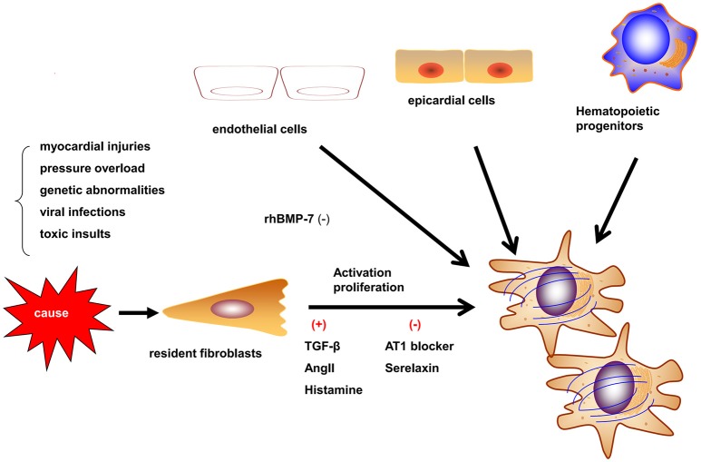
Myocardial fibrosis is an important part of cardiac remodeling that leads to heart failure and death. Myocardial fibrosis results from increased myofibroblast activity and excessive extracellular matrix deposition. Various cells and molecules are involved in this process, providing targets for potential drug therapies. Currently, the main detection methods of myocardial fibrosis rely on serum markers, cardiac magnetic resonance imaging, and endomyocardial biopsy. This review summarizes our current knowledge regarding the pathophysiology, quantitative assessment, and novel therapeutic strategies of myocardial fibrosis.
Keywords: biomarkers; extracelluar matrix; heart failure; late gadolinium enhancement; micro RNAs (miRNAs); mycardial fibrosis.
Figures

References
-
- Aoki T., Fukumoto Y., Sugimura K., Oikawa M., Satoh K., Nakano M., et al. (2011). Prognostic impact of myocardial interstitial fibrosis in non-ischemic heart failure. Comparison between preserved and reduced ejection fraction heart failure. Circ. J. 75, 2605–2613. 10.1253/circj.cj-11-0568 - DOI - PubMed
-
- aus dem Siepen F., Buss S. J., Messroghli D., Andre F., Lossnitzer D., Seitz S., et al. (2015). T1 mapping in dilated cardiomyopathy with cardiac magnetic resonance: quantification of diffuse myocardial fibrosis and comparison with endomyocardial biopsy. Eur. Heart J. Cardiovasc. Imaging 16, 210–216. 10.1093/ehjci/jeu183 - DOI - PubMed
Publication types
LinkOut - more resources
Full Text Sources
Other Literature Sources