Review
. 2017 Apr 24:8:134.
doi: 10.3389/fneur.2017.00134. eCollection 2017.

Ocular Tremor in Parkinson's Disease: Discussion, Debate, and Controversy

Affiliations
Review

Ocular Tremor in Parkinson's Disease: Discussion, Debate, and Controversy

Diego Kaski et al. Front Neurol. .

Abstract

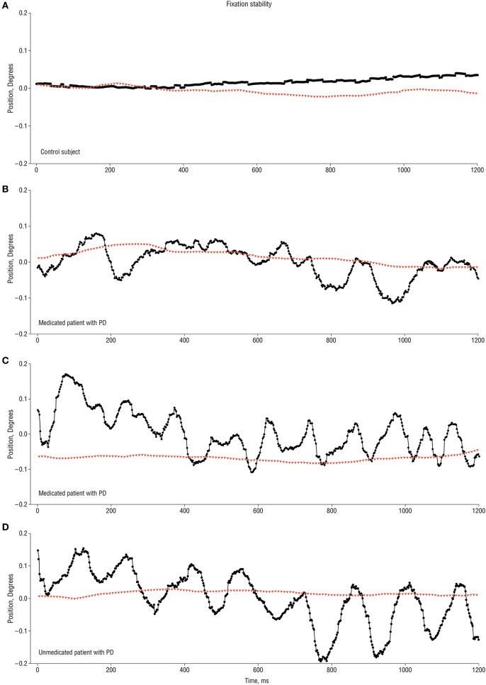
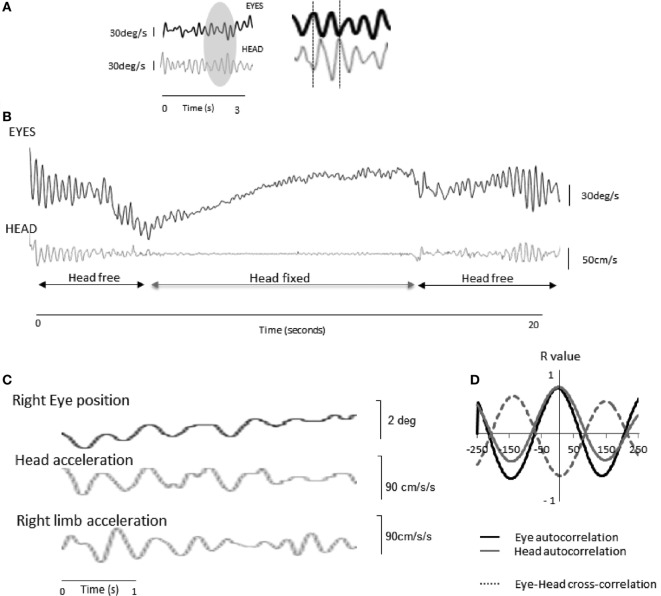
The identification of ocular tremor in a small cohort of patients with Parkinson's disease (PD) had lay somewhat dormant until the recent report of a pervasive ocular tremor as a universal finding in a large PD cohort that was, however, generally absent from a cohort of age-matched healthy subjects. The reported tremor had frequency characteristics similar to those of PD limb tremor, but the amplitude and frequency of the tremor did not correlate with clinical tremor ratings. Much controversy ensued as to the origin of such a tremor, and specifically as to whether a pervasive ocular tremor was a fundamental feature of PD, or rather a compensatory eye oscillation secondary to a transmitted head tremor, and thus a measure of a normal vestibulo-ocular reflex. In this mini review, we summarize some of the evidence for and against the case for a pervasive ocular tremor in PD and suggest future experiments that may help resolve these conflicting opinions.

Keywords: Parkinson’s disease; eye oscillations; head tremor; ocular tremor; vestibulo-ocular reflex.


Figures

Figure 1
Figure 1
1.2 s recording traces of ocular tremor in patients with Parkinson’s disease (PD) and healthy control [from Ref. (15) with permission]. (A) Eye movement recording from a control subject showing stable fixation (no ocular tremor). (B,C) Two different medicated PD patients showing eye oscillations of variable amplitude and approximately 6 Hz (B) and 10 Hz (C). (D) Ocular tremor in an unmedicated PD patient with a larger amplitude eye oscillation of approximately 10 Hz frequency. Note the absence of any head tremor in all traces. Black circles represent horizontal eye movements, with positive values indicting rightward eye movements, and red triangles indicating rotational head movement along the azimuth.
Figure 2
Figure 2
Eye and head oscillations in two patients with Parkinson’s disease (PD) [taken from Ref. (36) with permission]. (A) Patient 1—a 3 s recording of the eye (infrared video-oculography) and head (accelerometry) movements in a patient with PD with the head free.* To the right, a magnified view of the recordings reveals that the ocular oscillation is in antiphase to the head tremor. (B) When the head is physically restrained, the ocular tremor decreases in amplitude, indicating that the ocular tremor is intrinsically linked to the head tremor, and thus part of an intact VOR response. The ocular tremor is not abolished as the head can never be completely immobilized. (C) Raw oculographic, head, and limb tremor traces in a separate PD patient, without head restraint. The amplitude of the head and ocular tremor was smaller than in patient 1, in keeping with a smaller right limb tremor. Head tremor was not clinically visible in this patient. Head and limb oscillations were recorded from linear accelerometers. The eye and head traces appear in phase as a result of 180-degree phase shift between position (eye) and acceleration (head). (D) Autocorrelations for the eye and head traces and cross-correlation between the two signals over a 500-ms window for a fundamental frequency of 4.5 Hz. R values are shown in the y axis. *NB: In (A) and (B) head and eye traces are expressed in angular velocity units. Head acceleration values were, therefore, digitally integrated (linear acceleration to linear velocity), and corrected for eccentricity (tangential linear velocity to angular velocity, by taking into account occiput to head rotational axis distance, approximately 10 cm) using standard equations. As a leftward head rotation induces rightward occiput motion, the accelerometer trace has been inverted to correct for polarity. Finally, eye displacement recordings have been digitally differentiated (degrees to degrees/s).

Similar articles

Cited by

References

    1. Obeso JA, Rodríguez-Oroz MC, Benitez-Temino B, Blesa FJ, Guridi J, Marin C, et al. Functional organization of the basal ganglia: therapeutic implications for Parkinson’s disease. Mov Disord (2008) 23(Suppl 3):S548–59.10.1002/mds.22062 - DOI - PubMed
    1. Chaudhuri KR, Schapira AH. Non-motor symptoms of Parkinson’s disease: dopaminergic pathophysiology and treatment. Lancet Neurol (2009) 8:464–74.10.1016/S1474-4422(09)70068-7 - DOI - PubMed
    1. Martin WE, Loewenson RB, Resch JA, Baker AB. Parkinson’s disease. Clinical analysis of 100 patients. Neurology (1973) 23:783–90.10.1212/WNL.23.8.783 - DOI - PubMed
    1. Helmich RC, Hallett M, Deuschl G, Toni I, Bloem BR. Cerebral causes and consequences of Parkinsonian resting tremor: a tale of two circuits? Brain (2012) 135:3206–26.10.1093/brain/aws023 - DOI - PMC - PubMed
    1. Helmich RC, Janssen MJR, Oyen WJG, Bloem BR, Toni I. Pallidal dysfunction drives a cerebellothalamic circuit into Parkinson tremor. Ann Neurol (2011) 69:269–81.10.1002/ana.22361 - DOI - PubMed