. 2017 Apr 21:8:404.
doi: 10.3389/fimmu.2017.00404. eCollection 2017.

Anti-CD47 Antibody As a Targeted Therapeutic Agent for Human Lung Cancer and Cancer Stem Cells

Affiliations

Anti-CD47 Antibody As a Targeted Therapeutic Agent for Human Lung Cancer and Cancer Stem Cells

Liang Liu et al. Front Immunol. .

Abstract

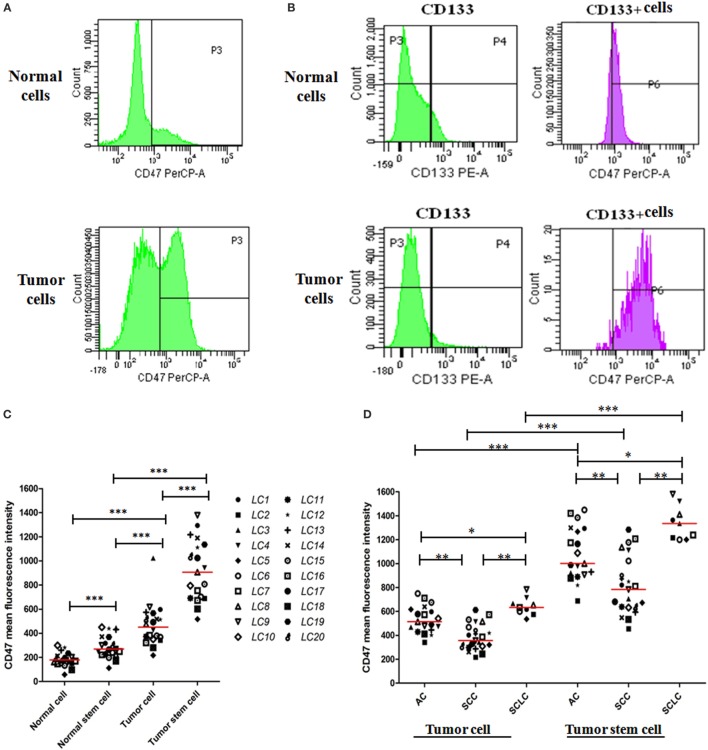
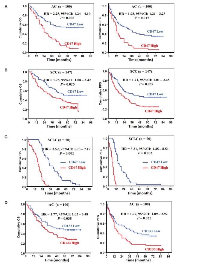
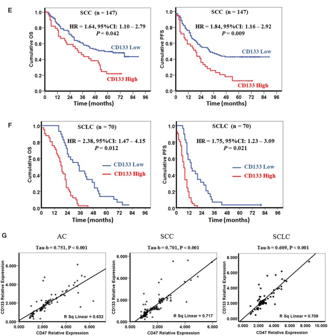
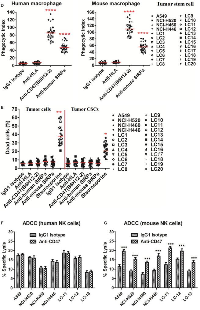
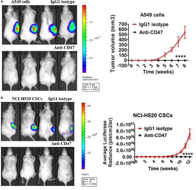
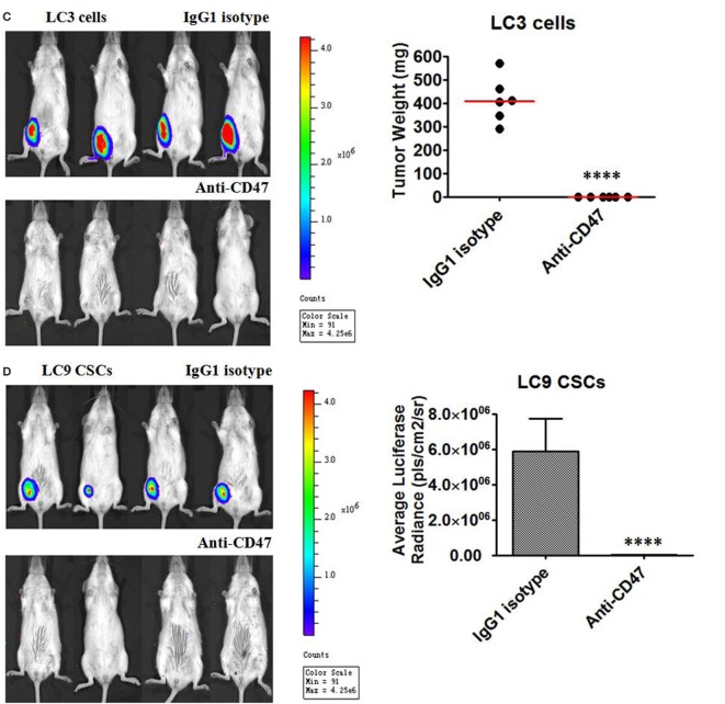
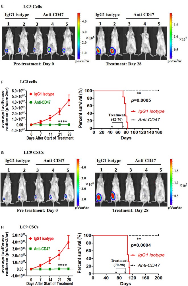
Accumulating evidence indicates that a small subset of cancer cells, termed the tumor-initiating cells or cancer stem cells (CSCs), construct a reservoir of self-sustaining cancer cells with the characteristic ability to self-renew and maintain the tumor mass. The CSCs play an important role in the tumor initiation, development, relapse, metastasis, and the ineffectiveness of conventional cancer therapies. CD47 is a ligand for signal-regulatory protein-α expressed on phagocytic cells and functions to inhibit phagocytosis. This study was to explore if the expression of CD47 is the mechanism used by lung cancer cells, especially CSCs, to escape phagocytosis in vitro and in vivo. Here, we selected CD133 as the marker for lung CSCs according to previous reports. We analyzed lung cancer and matched adjacent normal (non-tumor) tissue and revealed that CD47 is overexpressed on lung cancer cells, especially on lung CSCs. The mRNA expression levels of CD47 and CD133 correlated with a decreased probability of survival for multiple types of lung cancer. Blocking CD47 function with anti-CD47 antibodies enabled macrophage phagocytosis of lung cancer cells and lung CSCs. Anti-CD47 antibodies inhibited tumor growth in immunodeficient mouse xenotransplantation models established with lung cancer cells or lung CSCs and improved survival in tumor-bearing animals. These data indicate that CD47 is a valid target for cancer therapies, especially for anti-CSC therapies.

Keywords: CD47; antibody; cancer stem cells; human lung cancer; therapeutic agent.


Figures

Figure 1
Figure 1
CD47 expression is increased on lung cancer cells and lung cancer stem cells (CSCs) compared to their normal counterparts. (A) Levels of CD47 expression were detected on tumor cells and matched adjacent normal cells (normal cells) by flow cytometry (FACS). Cells analyzed were CD45, CD31, DAPI, and ESA+. (B) CD47 expression levels were detected on tumor stem cells CSCs and matched adjacent normal stem cells (normal stem cells) by FACS using the markers CD45, CD31, DAPI, ESA+, and CD133+ to selected cells for analysis. (C) CD47 expression on normal cells, normal stem cells, tumor cells, and CSCs was determined by FACS. Mean fluorescence intensity was normalized for cell size. Each data point represents a different patient sample: adenocarcinoma (AC) = 9, squamous cell carcinoma (SCC) = 9, small cell lung carcinoma (SCLC) = 2. P values were calculated using the paired-samples t-test. (D) CD47 expression across lung cancer subtypes including AC (n = 19), SCC (n = 21), and SCLC (n = 9) was determined as in (C). P values were calculated using the paired-samples t-test or the Two-independent samples test Mann–Whitney U model. *P < 0.05, **P < 0.01, and ***P < 0.001.
Figure 2
Figure 2
Increased CD47 and CD133 expressions correlate with a worse clinical prognosis. (A–C) CD47 mRNA level is an independent prognostic factor in lung cancers. Increased levels of CD47 mRNA were correlated with decreased probability of overall survival (OS) and progression-free survival (PFS) of patients with adenocarcinoma (AC) (A), squamous cell carcinoma (SCC) (B), and small cell lung carcinoma (SCLC) (C). (D–F) CD133 mRNA level is an independent prognostic factor in lung cancers. Prognostic impact of CD133 mRNA level is shown as the correlation with OS and PFS of patients with AC (D), SCC (E), and SCLC (F). P values were calculated using the Cox regression forward-LR model (A–F). (G) CD47 mRNA levels were positive correlation with the CD133 mRNA levels in patients with AC, SCC, and SCLC. P values were calculated using the Bivariate Correlations Kendall’s tau-b model.
Figure 2
Figure 2
Increased CD47 and CD133 expressions correlate with a worse clinical prognosis. (A–C) CD47 mRNA level is an independent prognostic factor in lung cancers. Increased levels of CD47 mRNA were correlated with decreased probability of overall survival (OS) and progression-free survival (PFS) of patients with adenocarcinoma (AC) (A), squamous cell carcinoma (SCC) (B), and small cell lung carcinoma (SCLC) (C). (D–F) CD133 mRNA level is an independent prognostic factor in lung cancers. Prognostic impact of CD133 mRNA level is shown as the correlation with OS and PFS of patients with AC (D), SCC (E), and SCLC (F). P values were calculated using the Cox regression forward-LR model (A–F). (G) CD47 mRNA levels were positive correlation with the CD133 mRNA levels in patients with AC, SCC, and SCLC. P values were calculated using the Bivariate Correlations Kendall’s tau-b model.
Figure 3
Figure 3
Blocking antibodies against CD47 enable phagocytosis of lung cancer cells and lung cancer stem cells (CSCs) by macrophages in vitro. (A,B) Carboxyfluoresceinsuccinimidyl ester (CFSE)-labeled lung cancer cells were incubated with human macrophages or mouse macrophages and the indicated antibodies and examined by immunofluorescence microscopy to detect phagocytosis. (A) Images from a representative lung cancer sample are shown. (B) Phagocytic indices of primary human lung cancer cells and lung cancer cell lines were determined using human (left) and mouse (right panel) macrophages. (C,D) CFSE-labeled lung CSCs were incubated with human macrophages or mouse macrophages as well as the indicated antibodies and examined by immunofluorescence microscopy to detect phagocytosis. (C) Images from a representative lung CSCs sample are shown. (D) Phagocytic indices of primary human lung CSCs and lung cancer cell lines CSCs were determined using human (left) and mouse (right panel) macrophages. (E) Antibody-induced apoptosis was tested by incubating lung cancer cells or lung CSCs with the indicated antibodies or staurosporine without macrophages and assessing the percentage of apoptotic and dead cells (% annexin V and/or PI positive). (F,G) Chromium release assays measuring ADCC were performed in triplicate with human (F) and mouse (G) at an effector:target ratio of 20:1, and percent specific lysis is reported. Antibodies were incubated at 10 μg/mL. P values were calculated using the two-independent samples test Mann–Whitney U model. *P < 0.05, **P < 0.01, ***P < 0.001, and ****P < 0.0001.
Figure 3
Figure 3
Blocking antibodies against CD47 enable phagocytosis of lung cancer cells and lung cancer stem cells (CSCs) by macrophages in vitro. (A,B) Carboxyfluoresceinsuccinimidyl ester (CFSE)-labeled lung cancer cells were incubated with human macrophages or mouse macrophages and the indicated antibodies and examined by immunofluorescence microscopy to detect phagocytosis. (A) Images from a representative lung cancer sample are shown. (B) Phagocytic indices of primary human lung cancer cells and lung cancer cell lines were determined using human (left) and mouse (right panel) macrophages. (C,D) CFSE-labeled lung CSCs were incubated with human macrophages or mouse macrophages as well as the indicated antibodies and examined by immunofluorescence microscopy to detect phagocytosis. (C) Images from a representative lung CSCs sample are shown. (D) Phagocytic indices of primary human lung CSCs and lung cancer cell lines CSCs were determined using human (left) and mouse (right panel) macrophages. (E) Antibody-induced apoptosis was tested by incubating lung cancer cells or lung CSCs with the indicated antibodies or staurosporine without macrophages and assessing the percentage of apoptotic and dead cells (% annexin V and/or PI positive). (F,G) Chromium release assays measuring ADCC were performed in triplicate with human (F) and mouse (G) at an effector:target ratio of 20:1, and percent specific lysis is reported. Antibodies were incubated at 10 μg/mL. P values were calculated using the two-independent samples test Mann–Whitney U model. *P < 0.05, **P < 0.01, ***P < 0.001, and ****P < 0.0001.
Figure 4
Figure 4
Ex vivo coating of lung cancer cells and lung cancer stem cells (CSCs) with an anti-CD47 antibody inhibits tumor engraftment. (A–D) Luciferase-expressing lung cancer cells [A549 cell line cells and primary LC3 tumor cells from a de novo small cell lung carcinoma (SCLC) patient of phase IIB] and lung CSCs [NCI-H520 cell line CSCs and primary LC9 CSCs from a de novo adenocarcinoma patient of phase IIIA] were precoated with IgG1 isotype control antibody or anti-human CD47 B6H12.2 antibody in vitro. NOD/SCID mice transplanted with the A549 cells (A), NCI-H520 CSCs (B), primary LC3 cells (C), or primary LC9 CSCs (D) were subject to bioluminescent imaging. Bioluminescence for A549 cells, NCI-H520 CSCs, LC3 cells, and LC9 CSCs engrafted mice was quantified (n = 6 per antibody condition). No tumor engraftment was observed in mice transplanted with anti-CD47-coated cells, in contrast to the 100% engraftment with IgG-coated cells (P < 0.0001), for all tested cells including A549 cells (A), NCI-H520 CSCs (B), LC3 cells (C), and LC9 CSCs (D). Data are represented as mean ± SD.
Figure 4
Figure 4
Ex vivo coating of lung cancer cells and lung cancer stem cells (CSCs) with an anti-CD47 antibody inhibits tumor engraftment. (A–D) Luciferase-expressing lung cancer cells [A549 cell line cells and primary LC3 tumor cells from a de novo small cell lung carcinoma (SCLC) patient of phase IIB] and lung CSCs [NCI-H520 cell line CSCs and primary LC9 CSCs from a de novo adenocarcinoma patient of phase IIIA] were precoated with IgG1 isotype control antibody or anti-human CD47 B6H12.2 antibody in vitro. NOD/SCID mice transplanted with the A549 cells (A), NCI-H520 CSCs (B), primary LC3 cells (C), or primary LC9 CSCs (D) were subject to bioluminescent imaging. Bioluminescence for A549 cells, NCI-H520 CSCs, LC3 cells, and LC9 CSCs engrafted mice was quantified (n = 6 per antibody condition). No tumor engraftment was observed in mice transplanted with anti-CD47-coated cells, in contrast to the 100% engraftment with IgG-coated cells (P < 0.0001), for all tested cells including A549 cells (A), NCI-H520 CSCs (B), LC3 cells (C), and LC9 CSCs (D). Data are represented as mean ± SD.
Figure 5
Figure 5
Therapy with anti-CD47 antibody eliminates lung cancer cells and lung cancer stem cells (CSCs) in xenotransplant models. (A) Luciferase-expressing A549 cells were transplanted subcutaneously into NOD/SCID mice. When palpable tumors (~100 mm3) formed, mice started to be treated with the IgG1 isotype antibody or anti-CD47 antibody (n = 6 per treatment group). Luciferase imaging of representative mice from each treatment group was shown before (day 0) and during (day 28) treatment (A). Average bioluminescence and improved survival were shown (B). (C) NOD/SCID mice were transplanted subcutaneously with luciferase-expressing NCI-H520 CSCs. When palpable tumors (~100 mm3) formed, mice started to be treated with the IgG1 isotype antibody or anti-CD47 antibody (n = 6 per treatment group). Luciferase imaging of representative mice from pretreatment and 28 days posttreatment were shown (C). Average bioluminescence and improved survival were shown (D). (E) NOD/SCID mice were transplanted subcutaneously with luciferase-expressing primary LC3 tumor cells. When palpable tumors (~100 mm3) formed, mice started to be treated with the IgG1 isotype antibody or anti-CD47 antibody (n = 6 per treatment group). Luciferase imaging of representative mice from pretreatment and 28 days posttreatment were shown (E). Average bioluminescence and improved survival were shown (F). (G) Luciferase-expressing primary LC9 CSCs were transplanted subcutaneously into the NOD/SCID mice. When palpable tumors (~100 mm3) formed, mice started to be treated with the IgG1 isotype antibody or anti-CD47 antibody (n = 6 per treatment group). Luciferase imaging of representative mice from pretreatment and 28 days posttreatment were shown (G). Average bioluminescence and improved survival were shown (H). ****P < 0.0001.
Figure 5
Figure 5
Therapy with anti-CD47 antibody eliminates lung cancer cells and lung cancer stem cells (CSCs) in xenotransplant models. (A) Luciferase-expressing A549 cells were transplanted subcutaneously into NOD/SCID mice. When palpable tumors (~100 mm3) formed, mice started to be treated with the IgG1 isotype antibody or anti-CD47 antibody (n = 6 per treatment group). Luciferase imaging of representative mice from each treatment group was shown before (day 0) and during (day 28) treatment (A). Average bioluminescence and improved survival were shown (B). (C) NOD/SCID mice were transplanted subcutaneously with luciferase-expressing NCI-H520 CSCs. When palpable tumors (~100 mm3) formed, mice started to be treated with the IgG1 isotype antibody or anti-CD47 antibody (n = 6 per treatment group). Luciferase imaging of representative mice from pretreatment and 28 days posttreatment were shown (C). Average bioluminescence and improved survival were shown (D). (E) NOD/SCID mice were transplanted subcutaneously with luciferase-expressing primary LC3 tumor cells. When palpable tumors (~100 mm3) formed, mice started to be treated with the IgG1 isotype antibody or anti-CD47 antibody (n = 6 per treatment group). Luciferase imaging of representative mice from pretreatment and 28 days posttreatment were shown (E). Average bioluminescence and improved survival were shown (F). (G) Luciferase-expressing primary LC9 CSCs were transplanted subcutaneously into the NOD/SCID mice. When palpable tumors (~100 mm3) formed, mice started to be treated with the IgG1 isotype antibody or anti-CD47 antibody (n = 6 per treatment group). Luciferase imaging of representative mice from pretreatment and 28 days posttreatment were shown (G). Average bioluminescence and improved survival were shown (H). ****P < 0.0001.
Figure 6
Figure 6
Anti-CD47 antibody exhibited no significant toxic effect except temporary white blood cell reduction and anemia. (A–D) Rat IgG2 isotype antibody or anti-mouse CD47 MIAP301 antibody was intraperitoneally injected into normal C57BL/6 mice at the dose of 400 µg daily from day 1 to day 14 (n = 4 per treatment group). These mice were followed up till day 28. White blood cell count (A), red blood cell count (B), and hemoglobin level (C) were temporarily decreased in anti-mouse CD47 group compared to IgG2 isotype group. Platelets count (D) has no difference between the two groups. (E–F) Mouse models were created as (A–D). Bone marrow from these mice was aspirated at day 0, day 14, and day 28 and indicated no effect of treatment on the frequency of LinKit+Sca+ hematopoietic stem cell (HSC) in the bone marrow. (E) Representative FACS plots are shown. (F) No differences in the percentage of HSC at day 0, day 14, and day 28 were observed with either control IgG or anti-mouse CD47.
Figure 6
Figure 6
Anti-CD47 antibody exhibited no significant toxic effect except temporary white blood cell reduction and anemia. (A–D) Rat IgG2 isotype antibody or anti-mouse CD47 MIAP301 antibody was intraperitoneally injected into normal C57BL/6 mice at the dose of 400 µg daily from day 1 to day 14 (n = 4 per treatment group). These mice were followed up till day 28. White blood cell count (A), red blood cell count (B), and hemoglobin level (C) were temporarily decreased in anti-mouse CD47 group compared to IgG2 isotype group. Platelets count (D) has no difference between the two groups. (E–F) Mouse models were created as (A–D). Bone marrow from these mice was aspirated at day 0, day 14, and day 28 and indicated no effect of treatment on the frequency of LinKit+Sca+ hematopoietic stem cell (HSC) in the bone marrow. (E) Representative FACS plots are shown. (F) No differences in the percentage of HSC at day 0, day 14, and day 28 were observed with either control IgG or anti-mouse CD47.

References

    1. Jordan CT, Guzman ML, Noble M. Cancer stem cells. N Engl J Med (2006) 355:1253–61. 10.1056/NEJMra061808 - DOI - PubMed
    1. Reya T, Morrison SJ, Clarke MF, Weissman IL. Stem cells, cancer, and cancer stem cells. Nature (2001) 414:105–11. 10.1038/35102167 - DOI - PubMed
    1. Pine SR, Ryan BM, Varticovski L, Robles AI, Harris CC. Microenvironmental modulation of asymmetric cell division in human lung cancer cells. Proc Natl Acad Sci U S A (2010) 107:2195–200. 10.1073/pnas.0909390107 - DOI - PMC - PubMed
    1. O’Brien CA, Kreso A, Jamieson CH. Cancer stem cells and self-renewal. Clin Cancer Res (2010) 16:3113–20. 10.1158/1078-0432.CCR-09-2824 - DOI - PubMed
    1. Bertolini G, Roz L, Perego P, Tortoreto M, Fontanella E, Gatti L, et al. Highly tumorigenic lung cancer CD133+ cells display stem-like features and are spared by cisplatin treatment. Proc Natl Acad Sci U S A (2009) 106:16281–6. 10.1073/pnas.0905653106 - DOI - PMC - PubMed