. 2017 Apr 21:8:459.
doi: 10.3389/fimmu.2017.00459. eCollection 2017.

Type I and Type III Interferons Display Different Dependency on Mitogen-Activated Protein Kinases to Mount an Antiviral State in the Human Gut

Affiliations

Type I and Type III Interferons Display Different Dependency on Mitogen-Activated Protein Kinases to Mount an Antiviral State in the Human Gut

Kalliopi Pervolaraki et al. Front Immunol. .

Abstract

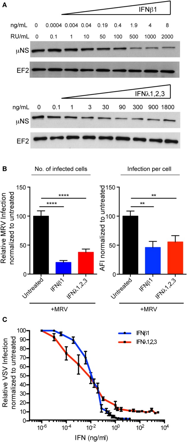
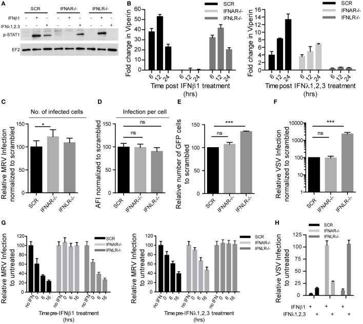
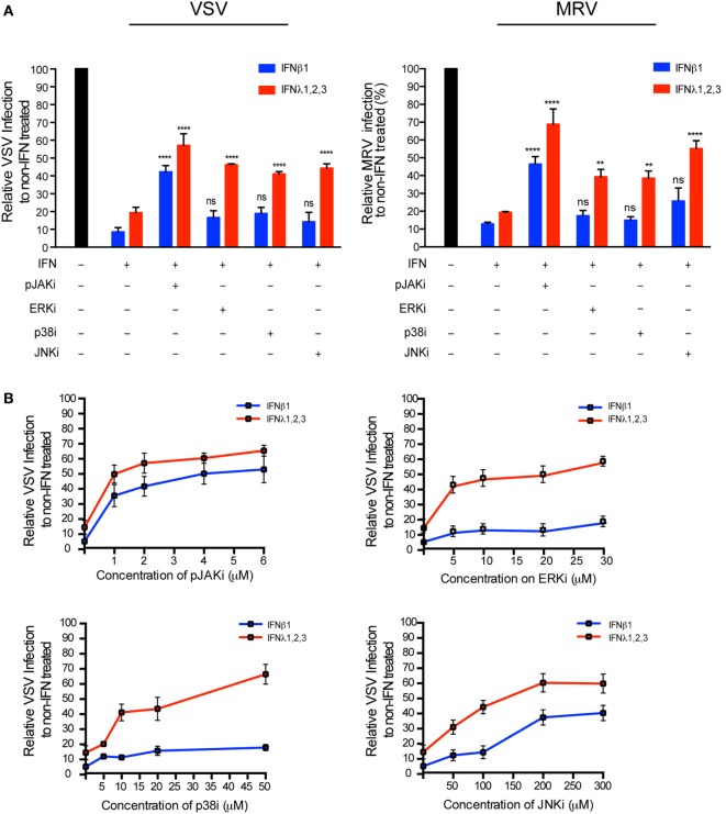
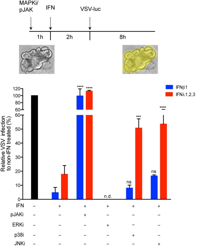
Intestinal epithelial cells (IECs) are constantly exposed to commensal flora and pathogen challenges. How IECs regulate their innate immune response to maintain gut homeostasis remains unclear. Interferons (IFNs) are cytokines produced during infections. While type I IFN receptors are ubiquitously expressed, type III IFN receptors are expressed only on epithelial cells. This epithelium specificity strongly suggests exclusive functions at epithelial surfaces, but the relative roles of type I and III IFNs in the establishment of an antiviral innate immune response in human IECs are not clearly defined. Here, we used mini-gut organoids to define the functions of types I and III IFNs to protect the human gut against viral infection. We show that primary non-transformed human IECs, upon viral challenge, upregulate the expression of both type I and type III IFNs at the transcriptional level but only secrete type III IFN in the supernatant. However, human IECs respond to both type I and type III IFNs by producing IFN-stimulated genes that in turn induce an antiviral state. Using genetic ablation of either type I or type III IFN receptors, we show that either IFN can independently restrict virus infection in human IECs. Importantly, we report, for the first time, differences in the mechanisms by which each IFN establishes the antiviral state. Contrary to type I IFN, the antiviral activity induced by type III IFN is strongly dependent on the mitogen-activated protein kinases signaling pathway, suggesting a pathway used by type III IFNs that non-redundantly contributes to the antiviral state. In conclusion, we demonstrate that human intestinal epithelial cells specifically regulate their innate immune response favoring type III IFN-mediated signaling, which allows for efficient protection against pathogens without producing excessive inflammation. Our results strongly suggest that type III IFN constitutes the frontline of antiviral response in the human gut. We propose that mucosal surfaces, particularly the gastrointestinal tract, have evolved to favor type III IFN-mediated response to pathogen infections as it allows for spatial segregation of signaling and moderate production of inflammatory signals which we propose are key to maintain gut homeostasis.

Keywords: antiviral immunity; human gut microbiota; interferon-lambda; interferon-β; intestinal epithelial cells; mitogen-activated protein kinases; mucosal immunity.


Figures

Figure 1
Figure 1
Infection of human mini-gut organoids with mammalian reovirus (MRV). (A) Human colon organoids were prepared according to methods. Representative images of human colon organoids grown over 10 days from intestinal crypts. (B) Five days post-differentiation, organoids were mock or MRV infected (multiplicity of infection = 0.5). 16 hpi, organoids were fixed, cryosectioned and immunostained for adherent junctions E-cadherin (E-cad), tight junctions (ZO-1), Goblet cells (Mucin-2), and Enteroendocrine cells (synaptophysin, Syn). (C) MRV-infected cells were detected using an antibody against the MRV non-structural protein μNS. Representative images are shown. White arrow indicates infected cells. Organoids were infected with MRV and virus replication was monitored by qRT-PCR over a timecourse of 24 h. Data represent the mean values of three independent experiments. Error bars indicate the SD.
Figure 2
Figure 2
Induction of immune response in human mini-gut organoids after mammalian reovirus (MRV) infection. Organoids were infected with MRV (multiplicity of infection = 0.5), quantitative real-time (qRT)-PCR and ELISA were used to detect (A) a time course of transcriptional upregulation of both type I interferons (IFNs) (β) and type III IFN (λ2/3) IFNs (B) 24 hpi the production and secretion of IFN proteins in the supernatant of infected organoids and (C) a time course of transcriptional upregulation of the IFN-stimulated genes Viperin and IFIT1. qRT-PCR data were normalized to TBP and HPRT1 (housekeeping genes) and are expressed relative to uninfected organoids at each time point. qRT-PCR data and ELISA data represent the mean values of three independent experiments. Error bars indicate the SD. The blue and red lines in (B) demarcate the limit of detection of our ELISA for type I and type III IFNs, respectively.
Figure 3
Figure 3
Both type I and type III interferons (IFNs) confer human mini-gut organoids protection against viral infection. (A) Colon organoids were treated with increasing concentrations of type I IFN (β) and type III IFN (λ1–3) IFN. Six hours posttreatment, organoids were harvested and the transcriptional upregulation of the IFN-stimulated genes Viperin (Vip) and Ifit1 was measured using qRT-PCR. Data were normalized to TBP and HPRT1. (B–E) Colon organoids were treated with type I IFN (β) (2,000 RU/mL equivalent 8 ng/mL) or type III IFN (λ1−3) (300 ng/mL) for 2.5 h prior to infection with mammalian reovirus (MRV) (multiplicity of infection = 0.5) for 16 h. (B) MRV-infected organoids were analyzed by μNS-specific immunofluorescence (green). The cells were stained against E-cadherin (red) and the nuclei were stained with Dapi (blue). Representative data from triplicate experiments are shown. White arrow indicates infected cells. (C) The fluorescence intensity of MRV μNS per organoid was measured and expressed relative to untreated organoids (set as 100). (D) MRV-infected organoids were analyzed for μNS production by Western blot. Actin was used as loading control. Production of μNS was quantified by densitometer. (E) The protective effect of type I or type III IFN was assayed by monitoring viral replication by qRT-PCR normalized to inoculum. Data represent the mean values of three independent experiments. Error bars indicate the SD. **P < 0.01, ***P < 0.001 (unpaired t-test).
Figure 4
Figure 4
Expression pattern of interferon (IFN) mRNA and protein in human intestinal epithelial cells upon viral infection (A). Relative quantification of type I IFN (β) and type III IFN (λ2/3) transcripts during the course of mammalian reovirus (MRV) (multiplicity of infection = 1) infection of T84 cells. Data are normalized to TBP and HPRT1 and are expressed relative to uninfected cells at each time point. (B) Quantification of type (IFNβ) and type III (IFN λ2/3) protein levels by ELISA in supernatants of uninfected or MRV-infected T84 cells. The blue and red dashed lines demarcate the limit of detection of our ELISA for type I and type III IFNs, respectively. n.d., not detectable. Data represent the mean values of three independent experiments. Error bars indicate the SD. ****P < 0.0001 (unpaired t-test).
Figure 5
Figure 5
Both type I and type III interferons (IFNs) mediate antiviral protection in human T84 cells. (A) T84 cells were pre-treated for 2.5 h with the indicated concentrations of type I IFN (β) and type III IFN (λ1–3) IFNs and then subsequently infected with mammalian reovirus (MRV) [multiplicity of infection (MOI) = 1]. Sixteen hours post-infection, the protective effect of type I or III IFN was assayed by immunoblotting for the viral non-structural protein μNS. EF-2 is used as a loading control. A representative immunoblot out of three independent experiments is shown. (B) T84 cells were treated with type I IFN (β) (2,000 RU/mL equivalent 8 ng/mL) or type III IFN (λ1−3) (300 ng/mL) for 2.5 h prior to infection with MRV for 16 h. MRV-infected cells were analyzed by μNS-specific immunofluorescence. (Left panel) The number of infected cells was quantified and is expressed relative to untreated cells (set to 100). (Right panel) MRV uNS staining intensity was measured to obtain the average fluorescent intensity per cell and is expressed relative to untreated cells (set to 100). Data represent the mean values of three independent experiments. (C) T84 cells were pre-treated with the indicated concentrations of type I or III IFNs for 2 h prior to infection with vesicular stomatis virus (VSV) expressing Firefly luciferase VSV expressing luciferase (MOI = 1). Viral replication was assayed by measuring the luciferase activity. For each sample luciferase activity was measured in triplicates and is expressed as the percentage of the activity present in VSV-infected cells without IFN treatment (set to 100). The mean value obtained from three independent experiments is plotted. Error bars indicate the SD. **P < 0.005, ****P < 0.0001 (unpaired t-test).
Figure 6
Figure 6
Type I and type III interferons (IFNs) independently confer intestinal epithelial cells antiviral protection. T84 IFNAR1 and IFNLR1 knockout cell lines were generated using the CRISPR/Cas9 system. (A) T84 cell lines were treated with type I IFN (β) (2,000 RU/mL equivalent 8 ng/mL) or type III IFN (λ1−3) (300 ng/mL) for 1 h and IFN signaling was measured by immunoblotting for pSTAT1 Y701. EF-2 is used as a loading control. A representative immunoblot out of three independent experiments is shown. (B) Same as (A), except that induction of IFN-stimulated genes was monitored by relative qRT-PCR quantification of Viperin at indicated times post-IFN treatment. Data were normalized to TBP and HPRT1 and are expressed relative to untreated cells of each time point. (C,D) T84 cell lines were infected with mammalian reovirus (MRV) for 16 h (multiplicity of infection (MOI) = 1) and MRV-infected cells were analyzed by μNS-specific immunofluorescence. (C) The number of infected cells is expressed relative to scramble control cells (set to 100). (D) MRV μNS staining intensity was measured to obtain the average fluorescence intensity per cell and expressed relative to scramble control cells (set to 100). (E) T84 cell lines were infected with vesicular stomatis virus (VSV)-GFP (MOI = 1) for 8 h and the number of VSV-infected cells were analyzed by FACS. The percentage of infected cells is expressed relative to scramble control cells (set to 100). (F) Same as (E), except that T84 cell lines were infected with VSV expressing luciferase (VSV-luc) (MOI = 1) and viral replication was assayed by measuring the luciferase activity. For each cell line luciferase activity was measured in triplicates and is expressed relative to scramble control cells (set to 100). (G) Same as (C), except that T84 cell lines were treated with type I IFN (β) (2,000 RU/mL equivalent 8 ng/mL) or type III IFN (λ1−3) (300 ng/mL) at indicated time points prior to infection with MRV. (H) Same as (F), except that T84 cell lines were treated with type I IFN (β) (2,000 RU/mL equivalent 8 ng/mL) or type III IFN (λ1−3) (300 ng/mL) for 2 h prior to infection with VSV-luc. Data (B–H) represent the mean values of three independent experiments. Error bars indicate the SD. *P < 0.05, ***P < 0.001, ns, not significant (unpaired t-test).
Figure 7
Figure 7
Type III interferons (IFNs) require mitogen-activated protein kinases for their antiviral response. (A) T84 cells were mock incubated (black bar) or pre-incubated for 30 min with 2 μM Pyridone 6 (pan-JAK inhibitor), 10 μM U0126 (ERK inhibitor), 10 μM SB202190 (p38 inhibitor), or 100 μM SP600125 (JNK inhibitor). Then, T84 cells were mock treated (black bar) or treated with type I IFN (β) (2,000 RU/mL equivalent 8 ng/mL) or type III IFN (λ1−3) (300 ng/mL) in the presence the inhibitor. Two hours post-IFN treatment cells were infected with a multiplicity of infection of 1 with VSV expressing luciferase (left panel) or mammalian reovirus (right panel). Viral replication was assayed by measuring the luciferase activity or by relative quantification of viral genome using qRT-PCR. Data were normalized to non-IFN-treated sample for each inhibitor (set to 100). (B) Same as (A), except T84 cells were pre-incubated with increasing concentrations of JAK or MAP kinase inhibitors prior to treatment with IFNs. The mean value obtained from three independent experiments, is plotted. Error bars indicate the SD. ****P < 0.0001, **P < 0.005, ns, not significant (unpaired t-test).
Figure 8
Figure 8
The antiviral activity of type III interferons (IFNs) strongly dependent on mitogen-activated protein kinases in the contact of primary human intestinal epithelial cells. Human colon organoids were mock incubated (black bar) or pre-incubated with 2 μM Pyridone 6 (pan-JAK inhibitor), 10 μM U0126 (ERK inhibitor), 10 μM M SB202190 (p38 inhibitor) and 100 μM SP600125 (JNK inhibitor). One hour posttreatment, organoids were mock treated (black bar) or co-treated with type I IFN (β) (2,000 RU/mL equivalent 8 ng/mL) or type III IFN (λ1−3) (300 ng/mL) for 2 h. Organoids were then infected with VSV expressing luciferase (multiplicity of infection = 1). Eight hpi, viral replication was assayed by measuring the luciferase activity. Data are normalized to non-IFN-treated sample for each inhibitor (set to 100). The mean value obtained from three independent experiments is plotted. Error bars indicate the SD. ****P < 0.0001, ***P < 0.001, ns, not significant (unpaired t-test), n.d. (not determined).

Similar articles

Cited by

References

    1. Pott J, Hornef M. Innate immune signalling at the intestinal epithelium in homeostasis and disease. EMBO Rep (2012) 13:684–98.10.1038/embor.2012.96 - DOI - PMC - PubMed
    1. Takeuchi O, Akira S. Innate immunity to virus infection. Immunol Rev (2008) 227(1):75–86.10.1111/j.1600-065X.2008.00737.x - DOI - PMC - PubMed
    1. Trinchieri G. Type I interferon: friend or foe? J Exp Med (2010) 207:2053–63.10.1084/jem.20101664 - DOI - PMC - PubMed
    1. Rubin DC, Shaker A, Levin MS. Chronic intestinal inflammation: inflammatory bowel disease and colitis-associated colon cancer. Front Immunol (2012) 3:107.10.3389/fimmu.2012.00107 - DOI - PMC - PubMed
    1. Kotenko SV, Gallagher G, Baurin VV, Lewis-Antes A, Shen M, Shah NK, et al. IFN-lambdas mediate antiviral protection through a distinct class II cytokine receptor complex. Nat Immunol (2003) 4:69–77.10.1038/ni875 - DOI - PubMed

LinkOut - more resources