Review
. 2017 Apr 24:8:475.
doi: 10.3389/fimmu.2017.00475. eCollection 2017.

The ST2/IL-33 Axis in Immune Cells during Inflammatory Diseases

Affiliations
Review

The ST2/IL-33 Axis in Immune Cells during Inflammatory Diseases

Brad Griesenauer et al. Front Immunol. .

Abstract

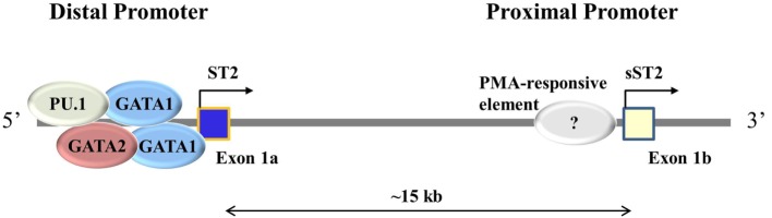
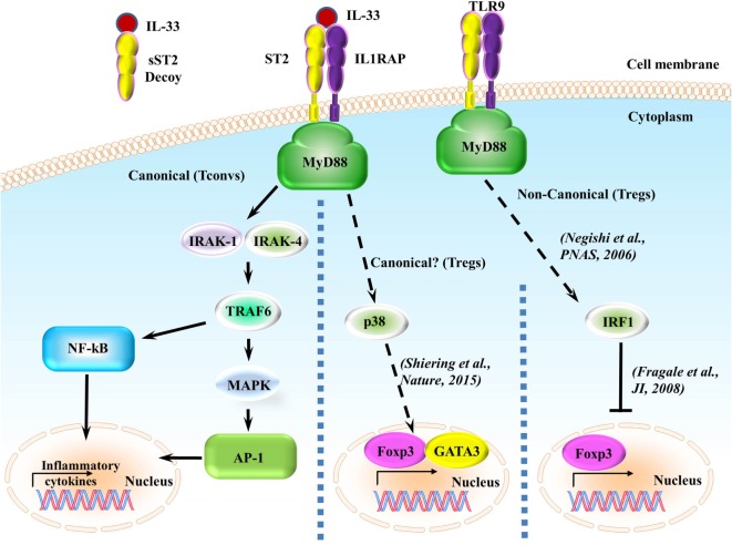
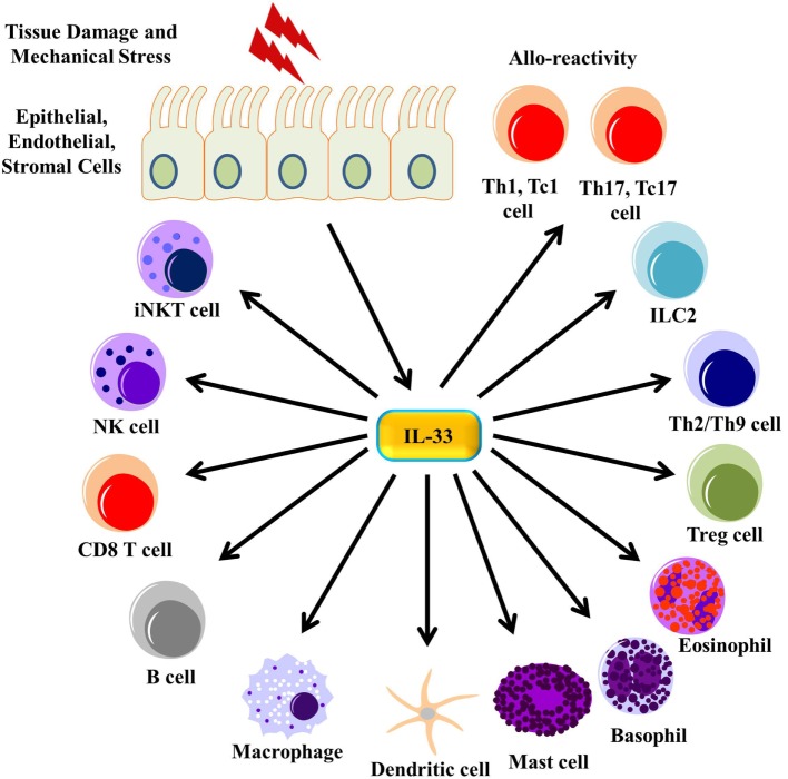
Il1rl1 (also known as ST2) is a member of the IL-1 superfamily, and its only known ligand is IL-33. ST2 exists in two forms as splice variants: a soluble form (sST2), which acts as a decoy receptor, sequesters free IL-33, and does not signal, and a membrane-bound form (ST2), which activates the MyD88/NF-κB signaling pathway to enhance mast cell, Th2, regulatory T cell (Treg), and innate lymphoid cell type 2 functions. sST2 levels are increased in patients with active inflammatory bowel disease, acute cardiac and small bowel transplant allograft rejection, colon and gastric cancers, gut mucosal damage during viral infection, pulmonary disease, heart disease, and graft-versus-host disease. Recently, sST2 has been shown to be secreted by intestinal pro-inflammatory T cells during gut inflammation; on the contrary, protective ST2-expressing Tregs are decreased, implicating that ST2/IL-33 signaling may play an important role in intestinal disease. This review will focus on what is known on its signaling during various inflammatory disease states and highlight potential avenues to intervene in ST2/IL-33 signaling as treatment options.

Keywords: IL-33; IL1RL1; ST2; cardiac diseases; graft-versus-host disease; lung diseases.


Figures

Figure 1
Figure 1
Different promoter usage dictates ST2 and sST2 expressions. ST2 consists of two main splice isoforms: ST2 and sST2. These isoforms are splice variants of each other regulated by alternative promoter bindings, the distal promoter for ST2, and the proximal promoter for sST2. Exon 1 varies between ST2 and sST2 depending on the promoter being bound. In immune cells, GATA1, GATA2, and PU.1 have been shown to bind to the distal promoter. The proximal promoter has not been well studied; it is thought that a PMA-responsive element induced sST2 transcription (37).
Figure 2
Figure 2
ST2/IL-33 signaling pathway. IL-33 either binds to the ST2/IL-1 receptor accessory protein (IL-1RAP) heterodimer, recruiting MyD88 to its intracellular domain, or the sST2 decoy receptor, which does not signal. MyD88 binding recruits IL-1R-associated kinase (IRAK) and TRAF6, leading to either the NF-κB or AP-1 pathway being activated. NF-κB and AP-1 activations promote inflammatory cytokine expressions. On regulatory T cells (Tregs), ST2/IL-33 signaling has been shown to promote Foxp3 and GATA3 expressions, while also promoting Treg function and expansion through enhancing TGF-β1-mediated differentiation though a p38-dependent mechanism. It has recently been shown that IFN regulatory factor (IRF) 1, which can be activated through MyD88 signaling, can inhibit Tregs by binding to the Foxp3 promoter and preventing Foxp3 transcription.
Figure 3
Figure 3
IL-33 signaling on immune cells. Tissue damage and mechanical stress to epithelial, endothelial, and stromal cells lead to the release of IL-33 from these cells. IL-33 then signals through many different immune cells, enhancing their function.
Figure 4
Figure 4
Pathogenesis of graft-versus-host disease (GVHD). The gut and other issues are damaged during irradiation or chemotherapy, leading to the release of various DAMPs, PAMPs, and cytokines, including IL-33. These DAMPs, PAMPs, and cytokines activate both host and donor antigen-presenting cells (APCs), which then activate the donor T cells. The APCs are also secreting various cytokines that promotes T cell differentiation toward type 1 and type 17 responses. These activated type 1 and type 17 T cells are able to secrete various pro-inflammatory cytokines, leading to apoptosis of healthy tissue, mainly in the gut, liver, and skin, which can be exacerbated by free IL-33. Furthermore, sST2 is produced by both type 1 and type 17 T cells, and while this may sequester free IL-33 from the type 1 and type 17 T cells, sST2 can also prevent the potential beneficial effects from ST2/IL-33 signaling in Th2 cells, Tregs, and lymphoid cell type 2 (ILC2s).

References

    1. Werenskiold AK, Hoffmann S, Klemenz R. Induction of a mitogen-responsive gene after expression of the Ha-ras oncogene in NIH 3T3 fibroblasts. Mol Cell Biol (1989) 9(11):5207–14.10.1128/MCB.9.11.5207 - DOI - PMC - PubMed
    1. Tominaga S. A putative protein of a growth specific cDNA from BALB/c-3T3 cells is highly similar to the extracellular portion of mouse interleukin 1 receptor. FEBS Lett (1989) 258(2):301–4.10.1016/0014-5793(89)81679-5 - DOI - PubMed
    1. Tominaga S-I, Ohta S, Tago K. Soluble form of the ST2 gene product exhibits growth promoting activity in NIH-3T3 cells. Biochem Biophys Rep (2016) 5:8–15.10.1016/j.bbrep.2015.11.020 - DOI - PMC - PubMed
    1. Meisel C, Bonhagen K, Lohning M, Coyle AJ, Gutierrez-Ramos JC, Radbruch A, et al. Regulation and function of T1/ST2 expression on CD4+ T cells: induction of type 2 cytokine production by T1/ST2 cross-linking. J Immunol (2001) 166(5):3143–50.10.4049/jimmunol.166.5.3143 - DOI - PubMed
    1. Schmitz J, Owyang A, Oldham E, Song Y, Murphy E, McClanahan TK, et al. IL-33, an interleukin-1-like cytokine that signals via the IL-1 receptor-related protein ST2 and induces T helper type 2-associated cytokines. Immunity (2005) 23(5):479–90.10.1016/j.immuni.2005.09.015 - DOI - PubMed