

Thoracic psammomatous spinal meningioma with osseous metaplasia: A very rare case report
- PMID: 28484549
- PMCID: PMC5409385
- DOI: 10.4103/1793-5482.150222
Thoracic psammomatous spinal meningioma with osseous metaplasia: A very rare case report
Abstract
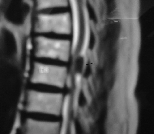
Thoracic spinal psammomatous meningioma is a rare subtype of meningioma. Among diverse types of mesenchymal differentiation, osseous metaplasia is found to be still rarer. We are presenting a new case of thoracic psammomatous spinal meningioma with osseous metaplasia in a middle aged female which that gives a sense of cancellous bone in the spinal canal. To conclude, meningiomas with osseous metaplasia are very rare tumors that complicate the surgical removal in certain cases. Ossification, if predicted prior to operation with computed tomography reconstruction, makes planning of removal easier. In our case, maintained cerebrospinal fluid spaces despite hard consistency of tumor made its removal easier once cerebrospinal fluid was drained. We have submitted this article because it is very rare and curable pathology and preoperative diagnosis helps in prevention of neurological injury during its excision.
Keywords: Ossified meningioma; osseous metaplasia; psammomatous meningioma.
Conflict of interest statement
There are no conflicts of interest.
Figures


References
-
- Louis DN, Scheithauer BW, Budka H. Meningiomas. In: Kleihues P, Cavenee WK, editors. World Health Organization Classification of Tumours, Pathology and Genetics of Tumours of the Nervous System. Lyon: IARC Press; 2000. pp. 176–84.
-
- Perry A, Louis DN, Scheithauer BW. Meningiomas. In: Louis DN, Ohgaki H, Wiestler OD, editors. World Health Organization Classification of Tumors of the Central Nervous System. Lyon: IARC Press; 2007. pp. 164–72.
-
- Liu CL, Lai PL, Jung SM, Liao CC. Thoracic ossified meningioma and osteoporotic burst fracture: Treatment with combined vertebroplasty and laminectomy without instrumentation: Case report. J Neurosurg Spine. 2006;4:256–9. - PubMed
-
- Naderi S, Yilmaz M, Canda T, Acar U. Ossified thoracic spinal meningioma in childhood: A case report and review of the literature. Clin Neurol Neurosurg. 2001;103:247–9. - PubMed
-
- Kato K, Chernov M, Urino T, Kasuya H, Kubo O, Isehi H, et al. Ossified frontosphenoorbital meningioma en plaque, mimicking extensive hyperostosis. Minim Invasive Neurosurg. 2008;51:237–9. - PubMed
Publication types
LinkOut - more resources
Full Text Sources
Other Literature Sources