

A rare case of dural arteriovenous fistula presenting as primary intraventricular hemorrhage
- PMID: 28484554
- PMCID: PMC5409390
- DOI: 10.4103/1793-5482.146399
A rare case of dural arteriovenous fistula presenting as primary intraventricular hemorrhage
Abstract
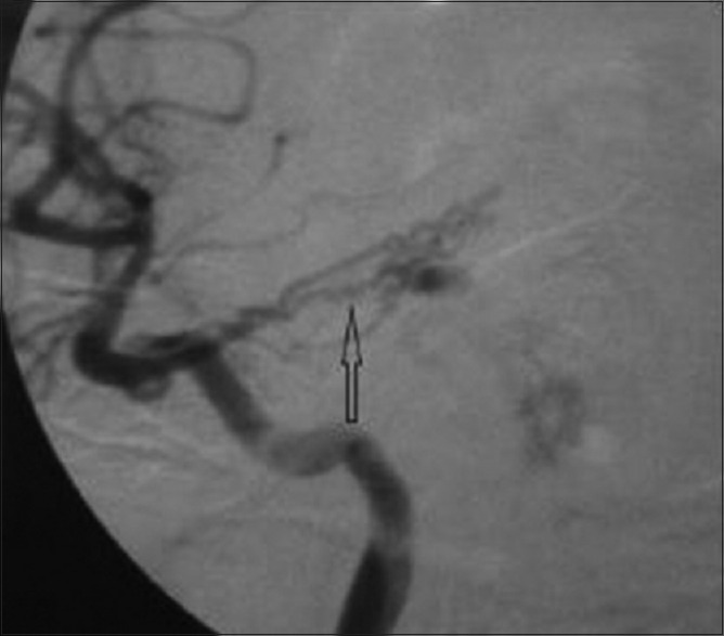
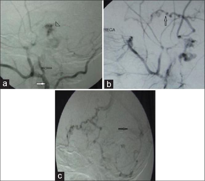
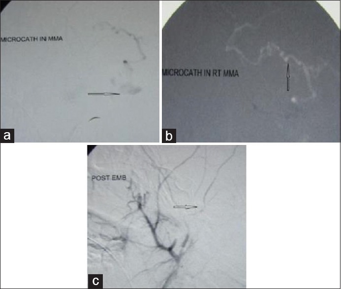
Primary intraventricular haemorrhage (PIVH) is rare. Dural arteriovenous fistula causing PIVH is extremely rare. We report a case of a 17 year old boy who presented with left hemiparesis, left lower motor neuron facial palsy and ataxia. His computed tomography head revealed primary intraventricular hemorrhage. Catheter super selective angiography revealed a dural arterio venous fistula with arterial feeder arising from the middle meningeal artery as well as from the inferior marginal tentorial artery. Glue injection led to successful disappearance of the fistula and eventual clinical recovery.
Keywords: Arterio-venous fistula; catheter angiography; cortico-venous reflux; primary intraventricular hemorrhage; venous sinuses.
Conflict of interest statement
There are no conflicts of interest.
Figures

Similar articles
-
Dural arteriovenous fistula causing primary intraventricular haemorrhage.Br J Radiol. 2008 Feb;81(962):e44-7. doi: 10.1259/bjr/25797982. Br J Radiol. 2008. PMID: 18238913
-
A dural arteriovenous fistula of the tentorium successfully treated by intravascular embolization.Surg Neurol. 1999 Sep;52(3):294-8. doi: 10.1016/s0090-3019(99)00076-2. Surg Neurol. 1999. PMID: 10511089
-
[A Case of Dural Arteriovenous Fistula Involving the Superior Sagittal Sinus that Presented as a Primary Intraventricular Hemorrhage].No Shinkei Geka. 2019 Aug;47(8):859-867. doi: 10.11477/mf.1436204037. No Shinkei Geka. 2019. PMID: 31477629 Japanese.
-
Isolated subdural hematoma secondary to Dural arteriovenous fistula: a case report and literature review.BMC Neurol. 2019 Mar 21;19(1):43. doi: 10.1186/s12883-019-1272-z. BMC Neurol. 2019. PMID: 30898107 Free PMC article. Review.
-
[A rare case of high cervical spinal cord dural arteriovenous fistula presenting with intracranial subarachnoid hemorrhage].No Shinkei Geka. 1994 Nov;22(11):1045-8. No Shinkei Geka. 1994. PMID: 7816174 Review. Japanese.
Cited by
-
Isolated intraventricular hemorrhage secondary to dural arteriovenous fistula.BMJ Case Rep. 2018 Feb 7;2018:bcr2017013643. doi: 10.1136/bcr-2017-013643. BMJ Case Rep. 2018. PMID: 29437717 Free PMC article.
-
Primary Intraventricular Hemorrhage Isolated in Cerebral Aqueduct Secondary to Dural Arteriovenous Fistula.J Vasc Interv Neurol. 2018 Nov;10(2):59-61. J Vasc Interv Neurol. 2018. PMID: 30746013 Free PMC article.
References
-
- Sanders E. A study of primary, immediate, or direct hemorrhage into the ventricles of the brain. Am J Med Sci. 1881;82:85–128.
-
- Darby DG, Donnan GA, Saling MA, Walsh KW, Bladin PF. Primary intraventricular hemorrhage: Clinical and neuropsychological findings in a prospective stroke series. Neurology. 1988;38:68–75. - PubMed
-
- Angelopoulos M, Gupta SR, Azat Kia B. Primary intraventricular hemorrhage in adults: Clinical features, risk factors, and outcome. Surg Neurol. 1995;44:433–6. - PubMed
-
- Brott T, Thalinger K, Hertzberg V. Hypertension as a risk factor for spontaneous intracerebral hemorrhage. Stroke. 1986;17:1078–83. - PubMed
-
- Giray S, Sen O, Sarica FB, Tufan K, Karatas M, Goksel BK, et al. Spontaneous primary intraventricular hemorrhage in adults: Clinical data, etiology and outcome. Turk Neurosurg. 2009;19:338–44. - PubMed
Publication types
LinkOut - more resources
Full Text Sources
Other Literature Sources