. 2017 Apr 5;4(4):160850.
doi: 10.1098/rsos.160850. eCollection 2017 Apr.

How do red blood cells know when to die?

Affiliations

How do red blood cells know when to die?

Clemente Fernandez Arias et al. R Soc Open Sci. .

Abstract

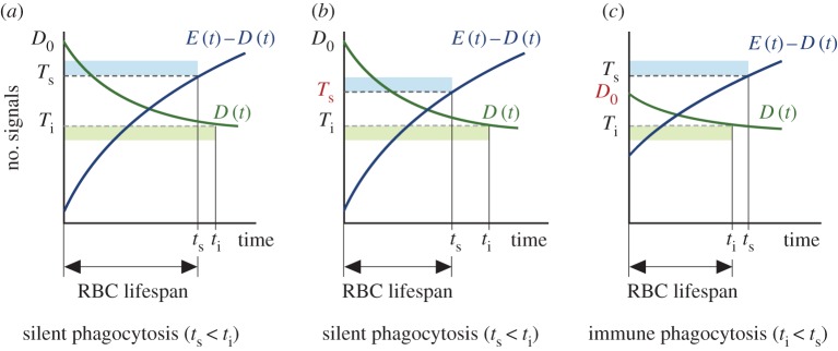
Human red blood cells (RBCs) are normally phagocytized by macrophages of splenic and hepatic sinusoids at 120 days of age. The destruction of RBCs is ultimately controlled by antagonist effects of phosphatidylserine (PS) and CD47 on the phagocytic activity of macrophages. In this work, we introduce a conceptual model that explains RBC lifespan as a consequence of the dynamics of these molecules. Specifically, we suggest that PS and CD47 define a molecular algorithm that sets the timing of RBC phagocytosis. We show that significant changes in RBC lifespan described in the literature can be explained as alternative outcomes of this algorithm when it is executed in different conditions of oxygen availability. The theoretical model introduced here provides a unified framework to understand a variety of empirical observations regarding RBC biology. It also highlights the role of RBC lifespan as a key element of RBC homeostasis.

Keywords: CD47; erythropoietin; neocytolysis; oxygen homeostasis; phosphatidylserine; red blood cell homeostasis.


Conflict of interest statement

The authors declare that they have no competing interests.

Figures

Figure 1.
Figure 1.
Rationale of the conceptual model of RBC lifespan determination. (a) Time evolution of membrane signals in a RBC according to empirical evidence (see points E1 and E2). (b) A RBC is phagocytized when the difference between ‘eat-me’ and ‘don’t-eat-me’ signals in its membrane attains a critical threshold (evidence E3). (c) An RBC can also be phagocytized if its level of ‘don’t-eat-me’ signals falls below a critical threshold (E4). (d) The conditions triggering RBC phagocytosis are mutually exclusive. In this example, the phagocytosis of the RBC occurs because the expression of ‘don’t-eat-me’ signals falls below a critical threshold (condition E4). (e,f) Different dynamics of membrane signals result in different lifespans (e) or in the RBC being phagocytized because it fulfils condition E3 before condition E4 (f).
Figure 2.
Figure 2.
Results of the mathematical model of RBC lifespan determination. (a) The dynamics of membrane signals as defined by equations (2.1)–(2.3) satisfy the qualitative constraints imposed by empirical evidence (E1–E4). Both the lifespan of the cell and how it is phagocytized (i.e. through the silent or the immune pathway) depend on the particular values of the model parameters. In this case, the difference between ‘eat-me’ and ‘don’t-eat-me’ signals is the first to reach its critical threshold (Ts), so that this cell is destroyed through the silent pathway at time ts (which sets its lifespan). (b) Changing the silent threshold (parameter Ts in the model) shortens the lifespan of the cell, but not the phagocytosis pathway. (c) By contrast, lower CD47 expression at the birth of the cell (parameter D0) both shortens the lifespan of the cell and changes the condition that triggers its phagocytosis (from silent to immune).
Figure 3.
Figure 3.
Potential mechanisms of RBC lifespan modulation. (a) Higher levels of oxidative stress are associated with higher rates of PS externalization. In agreement with empirical data, the model predicts an inverse correlation between the degree of OS and RBC lifespan. If the rate of PS externalization is above a critical value (β*) the curve of ts (time to reach the silent threshold) is below the curve of ti (time to attain the immune threshold). This implies that for high values of OS (β>β*) RBCs are phagocytized through the silent pathway. Only if β<β* are RBCs destroyed through the immune pathway, which can lead to anti-RBC autoimmunity. (b) Time evolution of the difference between ‘eat-me’ and ‘don’t-eat-me’ signals in the membrane of an RBC formed at time t1. The difference between membrane signals should reach the silent threshold at time t2, thereby causing the phagocytosis of the cell. Increasing the silent threshold delays the phagocytosis of the RBC until t3, thus extending its lifespan. (c) Neocytolysis. The figure shows the dynamics of the difference between ‘eat-me’ and ‘don’t-eat-me’ signals in the membrane of two RBCs that differ in the expression of membrane signals at birth. The first cell, formed at time t1, is phagocytized at time t4 after a normal lifespan. The second cell, born at time t2>t1 with a larger difference between ‘eat-me’ and ‘don’t-eat-me’ signals in its membrane attains the silent threshold much faster, so it is destroyed at time t3, before the first cell and after a much shorter lifespan. (d) According to our model, the lifespan of each RBC is directly correlated with the level of CD47 expressed in its membrane when it is formed. Low values of CD47 expression could explain short lifespans observed during neocytolysis. Furthermore, if the initial amount of CD47 falls below a critical level (the autoimmunity threshold), the immune phagocytosis occurs before the silent pathway (ti<ts). In this case, macrophages of the MPS phagocytize RBCs after very short lifespans and initiate anti-RBC autoimmune responses.
Figure 4.
Figure 4.
A theoretical model for the relationship between RBC lifespan and oxygen homeostasis. (a) Acclimation to environments with different partial pressures of oxygen, or clinical conditions that involve massive RBC loss such as malaria entail sharp fluctuations in the levels of plasma Epo (see text for references). (b) We hypothesize that Epo controls CD47 expression in newly formed RBCs, which in turn sets their expected lifespan (see equations (2.3)). In normal conditions both Epo and oxygen levels are at equilibrium, and mean RBC lifespan is around 120 days (0). Any variation in Epo, independently of its cause, changes the amount of CD47 in newly formed RBCs and hence its lifespan. From this perspective, a pronounced decrease in Epo suffices to account for the onset of neocytolysis observed in people returning to sea level after high-altitude acclimation or in malaria patients (labelled as −1 in the figure). Further drops of Epo can lead to autoimmunity (labelled as −2), which could explain the presence of auto-antibodies against host RBCs in malaria patients or in astronauts after space flights. See the text for further details.

References

    1. Wynn TA, Chawla A, Pollard JW. 2013. Macrophage biology in development, homeostasis and disease. Nature 496, 445–455. (doi:10.1038/nature12034) - DOI - PMC - PubMed
    1. Lang E, Qadri SM, Lang F. 2012. Killing me softly—suicidal erythrocyte death. Int. J. Biochem. Cell Biol. 44, 1236–1243. (doi:10.1016/j.biocel.2012.04.019) - DOI - PubMed
    1. Trial J, Rice L. 2004. Erythropoietin withdrawal leads to the destruction of young red cells at the endothelial-macrophage interface. Curr. Pharmaceut. Des. 10, 183–190. (doi:10.2174/1381612043453423) - DOI - PubMed
    1. Goodman JW, Smith LH. 1961. Erythrocyte life span in normal mice and in radiation bone marrow chimeras. Am. J. Physiol. 200, 764–770. - PubMed
    1. Horký J, Vácha J, Znojil V. 1977. Comparison of life span of erythrocytes in some inbred strains of mouse using 14C-labelled glycine. Physiol. Bohemoslovaca 27, 209–217. - PubMed

LinkOut - more resources