

Multiple huge epiphrenic esophageal diverticula with motility disease treated with video-assisted thoracoscopic and hand-assisted laparoscopic esophagectomy: a case report
- PMID: 28485002
- PMCID: PMC5422214
- DOI: 10.1186/s40792-017-0339-6
Multiple huge epiphrenic esophageal diverticula with motility disease treated with video-assisted thoracoscopic and hand-assisted laparoscopic esophagectomy: a case report
Abstract
Background: Epiphrenic esophageal diverticulum is a rare condition that is often associated with a concomitant esophageal motor disorder. Some patients have the chief complaints of swallowing difficulty and gastroesophageal reflux; traditionally, such diverticula have been resected via right thoracotomy. Here, we describe a case with huge multiple epiphrenic diverticula with motility disorder, which were successfully resected using a video-assisted thoracic and laparoscopic procedure.
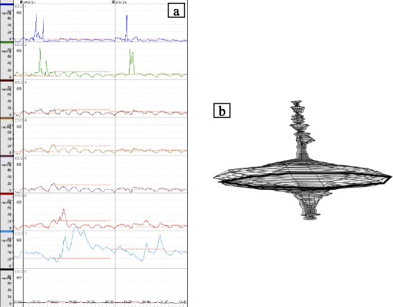
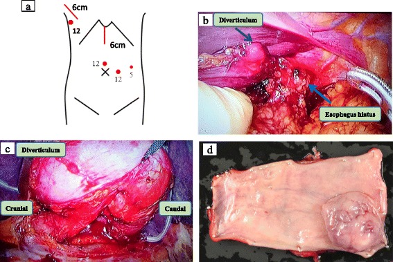
Case presentation: A 63-year-old man was admitted due to dysphagia, heartburn, and vomiting. An esophagogram demonstrated an S-shaped lower esophagus with multiple epiphrenic diverticula (75 × 55 mm and 30 × 30 mm) and obstruction by the lower esophageal sphincter (LES). Esophageal manometry showed normal peristaltic contractions in the esophageal body, whereas the LES pressure was high (98.6 mmHg). The pressure vector volume of LES was 23,972 mmHg2 cm. Based on these findings, we diagnosed huge multiple epiphrenic diverticula with a hypertensive lower esophageal sphincter and judged that resection might be required. We performed lower esophagectomy with gastric conduit reconstruction using a video-assisted thoracic and hand-assisted laparoscopic procedure. The postoperative course was uneventful, and the esophagogram demonstrated good passage, with no leakage, stenosis, or diverticula.
Conclusions: The most common causes of mid-esophageal and epiphrenic diverticula are motility disorders of the esophageal body; appropriate treatment should be considered based on the morphological and motility findings.
Keywords: Epiphrenic esophageal diverticulum; Esophageal motility; Hypertensive lower esophageal sphincter; Video-assisted thoracic surgery.
Figures

References
LinkOut - more resources
Full Text Sources
Other Literature Sources
