

Monitoring Changes in Hepatic Venous Velocities Flow after a Fluid Challenge Can Identify Shock Patients Who Lack Fluid Responsiveness
- PMID: 28485321
- PMCID: PMC5443027
- DOI: 10.4103/0366-6999.205848
Monitoring Changes in Hepatic Venous Velocities Flow after a Fluid Challenge Can Identify Shock Patients Who Lack Fluid Responsiveness
Abstract
Background: Evaluating the hemodynamic status and predicting fluid responsiveness are important in critical ultrasound assessment of shock patients. Transthoracic echocardiography with noninvasive diagnostic parameters allows the assessment of volume responsiveness. This study aimed to assess the hemodynamic changes in the liver and systemic hemodynamic changes during fluid challenge and during passive leg raising (PLR) by measuring hepatic venous flow (HVF) velocity.
Methods: This is an open-label study in a tertiary teaching hospital. Shock patients with hypoperfusion who required fluid challenge were selected for the study. Patients <18 years old and those with contraindications to PLR were excluded from the study. Baseline values were measured, PLR tests were performed, and 500 ml of saline was infused over 30 min. Parameters associated with cardiac output (CO) in the left ventricular outflow tract were measured using the Doppler method. In addition, HVF velocity and right ventricular function parameters were determined.
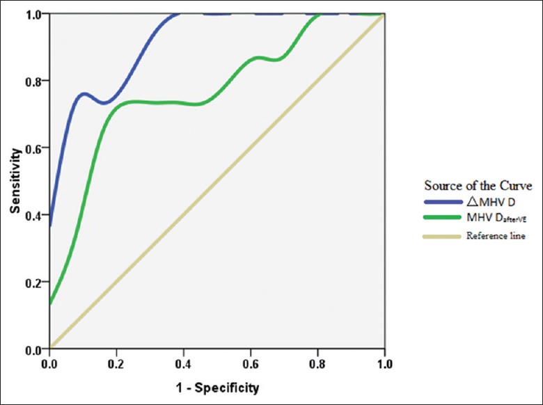
Results: Middle hepatic venous (MHV) S-wave velocity was positively correlated in all patients with CO at baseline (r = 0.706, P< 0.01) and after volume expansion (r = 0.524, P= 0.003). CO was also significantly correlated with MHV S-wave velocity in responders (r = 0.608, P< 0.01). During PLR, however, hepatic venous S-wave velocity did not correlate with CO. For the parameter ΔMHV D (increase in change in MHV D-wave velocity after volume expansion), defined as (MHV DafterVE - MHV DBaseline)/MHV DBaseline× 100%, >21% indicated no fluid responsiveness, with a sensitivity of 100%, a specificity of 71.2%, and an area under the receiver operating characteristic curve of 0.918.
Conclusions: During fluid expansion, hepatic venous S-wave velocity can be used to monitor CO, whether or not it is increasing. ΔMHV D ≥21% indicated a lack of fluid responsiveness, thus helping to decide when to stop infusions.
Conflict of interest statement
There are no conflicts of interest.
Figures


References
-
- Zhang Z, Xu X, Ye S, Xu L. Ultrasonographic measurement of the respiratory variation in the inferior vena cava diameter is predictive of fluid responsiveness in critically ill patients: Systematic review and meta-analysis. Ultrasound Med Biol. 2014;40:845–53. doi: 10.1016/j.ultrasmedbio.2013.12.010. - PubMed
-
- Guyton AC. Determination of cardiac output by equating venous return curves with cardiac response curves. Physiol Rev. 1955;35:123–9. - PubMed
-
- Scheinfeld MH, Bilali A, Koenigsberg M. Understanding the spectral Doppler waveform of the hepatic veins in health and disease. Radiographics. 2009;29:2081–98. doi: 10.1148/rg.297095715. - PubMed
-
- Mahjoub Y, Touzeau J, Airapetian N, Lorne E, Hijazi M, Zogheib E, et al. The passive leg-raising maneuver cannot accurately predict fluid responsiveness in patients with intra-abdominal hypertension. Crit Care Med. 2010;38:1824–9. doi: 10.1097/CCM.0b013e3181eb3c21. - PubMed
MeSH terms
LinkOut - more resources
Full Text Sources
Other Literature Sources
Medical
