

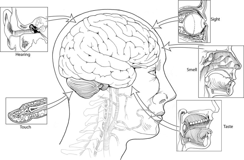
Overview of Neuropeptides: Awakening the Senses?
- PMID: 28485842
- PMCID: PMC5424629
- DOI: 10.1111/head.13084
Overview of Neuropeptides: Awakening the Senses?
Abstract
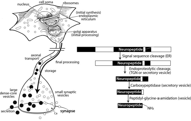
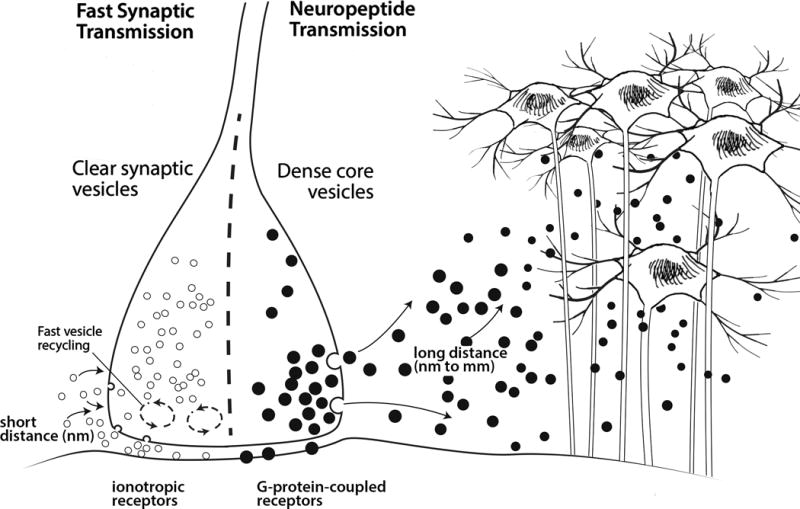
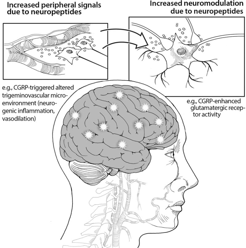
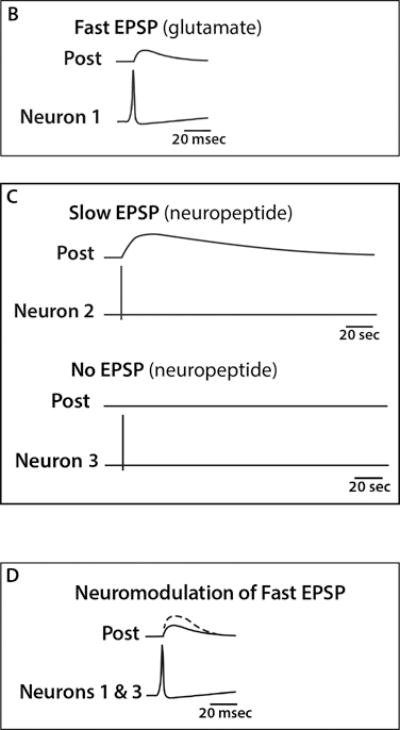
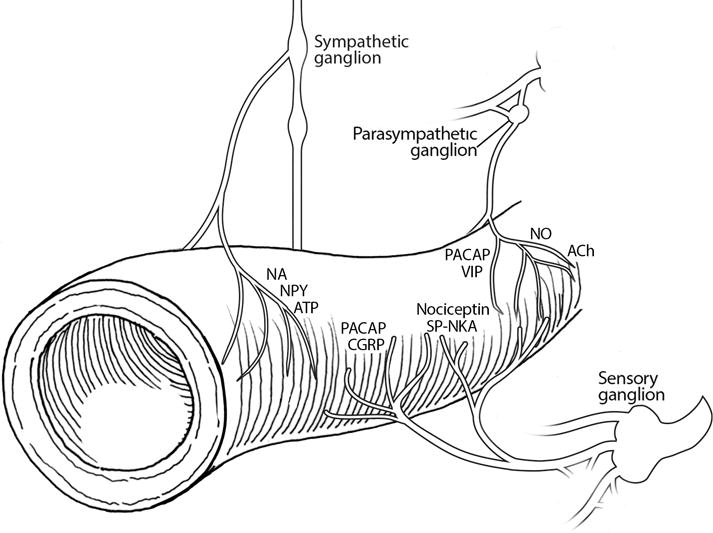
Humans have a diverse collection of neuropeptides that can influence a multitude of activities. There are now over 100 known neuropeptides and probably many more yet to be identified from the over 1000 predicted peptides encoded in the genome. While diverse, peptides generally share three common characteristics: (1) post-translational processing and release from vesicles, (2) activation of cell-surface receptors over a relatively large distance, and (3) modulation of target cells that are often in the brain and periphery. Within the brain, neuropeptides can modulate the activity of co-released neurotransmitters to either increase or decrease the strength of synaptic signaling. Within the periphery, neuropeptides can function similar to peptide hormones and modulate nearly all bodily functions. Given the clear involvement of the neuropeptide CGRP in migraine and the emerging evidence for other peptides, it seems likely that neuropeptides may help "awaken" the senses and contribute to the heightened sensory state of migraine.
Keywords: CGRP; neuromodulation; neuropeptide.
© 2017 American Headache Society.
Conflict of interest statement
Figures

Similar articles
-
Neuropeptides in sensory signal processing.Cell Tissue Res. 2019 Jan;375(1):217-225. doi: 10.1007/s00441-018-2946-3. Epub 2018 Oct 30. Cell Tissue Res. 2019. PMID: 30377783 Review.
-
Neuropeptides and neuropeptide receptors: drug targets, and peptide and non-peptide ligands: a tribute to Prof. Dieter Seebach.Chem Biodivers. 2012 Nov;9(11):2367-87. doi: 10.1002/cbdv.201200288. Chem Biodivers. 2012. PMID: 23161624 Review.
-
What are neuropeptides?Methods Mol Biol. 2011;789:1-36. doi: 10.1007/978-1-61779-310-3_1. Methods Mol Biol. 2011. PMID: 21922398
-
Neuropeptides as a Marker for Chronic Headache.Curr Pain Headache Rep. 2017 Apr;21(4):18. doi: 10.1007/s11916-017-0618-8. Curr Pain Headache Rep. 2017. PMID: 28281109 Review.
-
Neuropeptides from concept to online database www.neuropeptides.nl.Eur J Pharmacol. 2010 Jan 10;626(1):27-48. doi: 10.1016/j.ejphar.2009.10.015. Epub 2009 Oct 27. Eur J Pharmacol. 2010. PMID: 19837055 Review.
Cited by
-
Mechanism of Action of OnabotulinumtoxinA in Chronic Migraine: A Narrative Review.Headache. 2020 Jul;60(7):1259-1272. doi: 10.1111/head.13849. Epub 2020 Jun 30. Headache. 2020. PMID: 32602955 Free PMC article. Review.
-
Human iN neuronal model of schizophrenia displays dysregulation of chromogranin B and related neuropeptide transmitter signatures.Mol Psychiatry. 2024 May;29(5):1440-1449. doi: 10.1038/s41380-024-02422-x. Epub 2024 Feb 2. Mol Psychiatry. 2024. PMID: 38302561 Free PMC article.
-
Alternative Pharmacological Strategies for the Treatment of Alzheimer's Disease: Focus on Neuromodulator Function.Biomedicines. 2022 Nov 28;10(12):3064. doi: 10.3390/biomedicines10123064. Biomedicines. 2022. PMID: 36551821 Free PMC article. Review.
-
The Local Neuropeptide System of Keratinocytes.Biomedicines. 2021 Dec 7;9(12):1854. doi: 10.3390/biomedicines9121854. Biomedicines. 2021. PMID: 34944669 Free PMC article. Review.
-
A Global Review on Short Peptides: Frontiers and Perspectives.Molecules. 2021 Jan 15;26(2):430. doi: 10.3390/molecules26020430. Molecules. 2021. PMID: 33467522 Free PMC article. Review.
References
-
- Burbach JP. What are neuropeptides? Methods Mol Biol. 2011;789:1–36. - PubMed
-
- Pert C. Molecules of emotion: The science behind mind-body medicine. New York: Scribner; 1997.
-
- ScribnerHUGO gene nomenclature committee. Neuropeptide database. Human Genome Organization; 2017.
Publication types
MeSH terms
Substances
Grants and funding
LinkOut - more resources
Full Text Sources
Other Literature Sources
Research Materials
