Review
. 2017 Aug;90(1077):20170197.
doi: 10.1259/bjr.20170197. Epub 2017 May 25.

Breast metastases from extramammary malignancies: multimodality imaging aspects

Affiliations
Review

Breast metastases from extramammary malignancies: multimodality imaging aspects

Almir G V Bitencourt et al. Br J Radiol. 2017 Aug.

Abstract

Breast metastases from extramammary cancers are rare and usually related to poor prognosis. The extramammary tumours most frequently exhibiting breast metastases are melanoma, lymphomas, ovarian cancer, lung and neuroendocrine tumours, and sarcomas. Owing to the lack of reliable and specific clinical or radiological signs for the diagnosis of breast metastases, a combination of techniques is needed to differentiate these lesions from primary breast carcinoma or even benign breast lesions. Multiple imaging methods may be used to evaluate these patients, including mammography, ultrasound, MRI, CT and positron emission tomography CT. Clinical and imaging manifestations are varied, depend on the form of dissemination of the disease and may mimic primary benign and malignant breast lesions. Haematologically disseminated metastases often develop as a circumscribed mass, whereas lymphatic dissemination often presents as diffuse breast oedema and skin thickening. Unlike primary carcinomas, breast metastases generally do not have spiculated margins, skin or nipple retraction. Microlobulated or indistinct margins may be present in some cases. Although calcifications are not frequently present in metastatic lesions, they occur more commonly in patients with ovarian cancer. Although rare, secondary malignant neoplasms should be considered in the differential diagnosis of breast lesions, in the appropriate clinical setting. Knowledge of the most common imaging features can help to provide the correct diagnosis and adequate therapeutic planning.


Figures
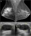
Figure 1.
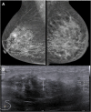
Figure 1.
A 73-year-old female patient with a palpable lump in the right breast. Mammography showed a hyperdense mass with obscured margins on upper-outer quadrant to the right breast and enlarged axillary lymph nodes (a: mediolateral oblique views). Ultrasound showed an oval hypoechoic mass with circumscribed margins (b). Biopsy showed lymphoid cells with vesicular nuclei and high mitotic index, compatible with non-Hodgkin lymphoma.
Figure 2.
Figure 2.
A 50-year-old female patient treated for non-Hodgkin's lymphoma 2 years earlier with complete remission, presented with fever and diffuse hyperaemia of the right breast (a), without response to systemic antibiotics. Ultrasound showed heterogeneous mass with indistinct margins (b). Contrast-enhanced chest CT showed a heterogeneous mass with associated oedema (c) and intense fluorine-18-labelled fludeoxyglucose uptake on positron emission tomography/CT (d). Biopsy revealed anaplastic large-cell lymphoma.
Figure 3.
Figure 3.
A 52-year-old female patient with history of melanoma on the left thigh 2 years earlier complained of palpable and painful lump in the right breast. Mammography (a: mediolateral oblique view) showed a round circumscribed mass in the lower-outer quadrant of the right breast (arrow). Breast ultrasound showed a hypoechoic mass with indistinct margins and mild rim vascularity on colour Doppler (b). MRI showed an irregular mass with heterogeneous enhancement (arrow) with washout kinetic curve (c). Percutaneous biopsy was compatible with metastatic melanoma.
Figure 4.
Figure 4.
A 48-year-old female patient with previous melanoma on the thumb. Mammography (a: mediolateral oblique view) showed an oval circumscribed mass in the upper-outer quadrant of the left breast. Breast ultrasound showed an oval circumscribed mass with heterogeneous echo pattern (b). Percutaneous biopsy showed metastatic melanoma.
Figure 5.
Figure 5.
A 39-year-old female patient submitted to surgical resection of a retroperitoneal leiomyosarcoma 1 year earlier, presented a mass in the right breast on screening tests. Mammography identified a round circumscribed mass (arrow) in the lower-inner quadrant of the right breast (a: mediolateral oblique view). Ultrasound showed a round, circumscribed, complex solid and cystic mass with internal vascularity on colour Doppler (b). Percutaneous biopsy of the lesion confirmed metastatic leiomyosarcoma.
Figure 6.
Figure 6.
A 45-year-old female patient under treatment for metastatic neuroendocrine tumour presented a new small mass on routine breast ultrasound. Breast ultrasound showed a small hypoechoic circumscribed mass (arrows) in the upper quadrants of the left breast (a), which was also identified on MRI (b). The small mass on the left breast (arrow) showed increased uptake of gallium-68 DOTATATE on positron emission tomography-CT (c), which suggest a well-differentiated neuroendocrine metastasis.
Figure 7.
Figure 7.
A 77-year-old female patient with prior serous ovarian adenocarcinoma presented an oval mass with pleomorphic calcifications (arrow) at mammography (a). Breast ultrasound also showed calcifications within an oval hypoechoic mass (b). Percutaneous biopsy was positive for metastatic ovarian carcinoma.
Figure 8.
Figure 8.
A 64-year-old female patient with lung adenocarcinoma presented with oedema of the left breast. Mammography showed skin and trabecular thickening, associated with axillary adenopathy (a). Breast ultrasound showed an ill-defined hypoechoic lesion in the upper quadrants of the left breast (b) and abnormal axillary lymph nodes. Ultrasound-guided biopsy confirmed metastatic lung adenocarcinoma in the breast.

References

    1. Bartella L, Kaye J, Perry NM, Malhotra A, Evans D, Ryan D, et al. Metastases to the breast revisited: radiological–histopathological correlation. Clin Radiol 2003; 58: 524–31. doi: https://doi.org/10.1016/s0009-9260(03)00068-0 - DOI - PubMed
    1. Buisman FE, van Gelder L, Menke-Pluijmers MBE, Bisschops BHC, Plaisier PW, Westenend PJ. Non-primary breast malignancies: a single institution's experience of a diagnostic challenge with important therapeutic consequences—a retrospective study. World J Surg Oncol 2016; 14: 166. doi: https://doi.org/10.1186/s12957-016-0915-4 - DOI - PMC - PubMed
    1. Sippo DA, Kulkarni K, Carlo PD, Lee B, Eisner D, Cimino-Mathews A, et al. Metastatic disease to the breast from extramammary malignancies: a multimodality pictorial review. Curr Probl Diagn Radiol 2016; 45: 225–32. doi: https://doi.org/10.1067/j.cpradiol.2015.07.001 - DOI - PubMed
    1. Abbas J, Wienke A, Spielmann RP, Bach AG, Surov A. Intramammary metastases: comparison of mammographic and ultrasound features. Eur J Radiol 2013; 82: 1423–30. doi: https://doi.org/10.1016/j.ejrad.2013.04.032 - DOI - PubMed
    1. Lee SK, Kim WW, Kim SH, Hur SM, Kim S, Choi JH, et al. Characteristics of metastasis in the breast from extramammary malignancies. J Surg Oncol 2010; 101: 137–40. doi: https://doi.org/10.1002/jso.21453 - DOI - PubMed

MeSH terms