. 2018 Jan;24(1-2):135-144.
doi: 10.1089/ten.TEA.2017.0044. Epub 2017 Jun 13.

Role of Bone Marrow Mononuclear Cell Seeding for Nanofiber Vascular Grafts

Affiliations

Role of Bone Marrow Mononuclear Cell Seeding for Nanofiber Vascular Grafts

Takuma Fukunishi et al. Tissue Eng Part A. 2018 Jan.

Abstract

Objective: Electrospinning is a promising technology that provides biodegradable nanofiber scaffolds for cardiovascular tissue engineering. However, success with these materials has been limited, and the optimal combination of scaffold parameters for a tissue-engineered vascular graft (TEVG) remains elusive. The purpose of the present study is to evaluate the effect of bone marrow mononuclear cell (BM-MNC) seeding in electrospun scaffolds to support the rational design of optimized TEVGs.

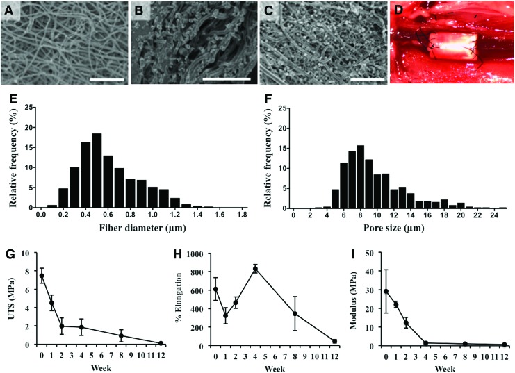
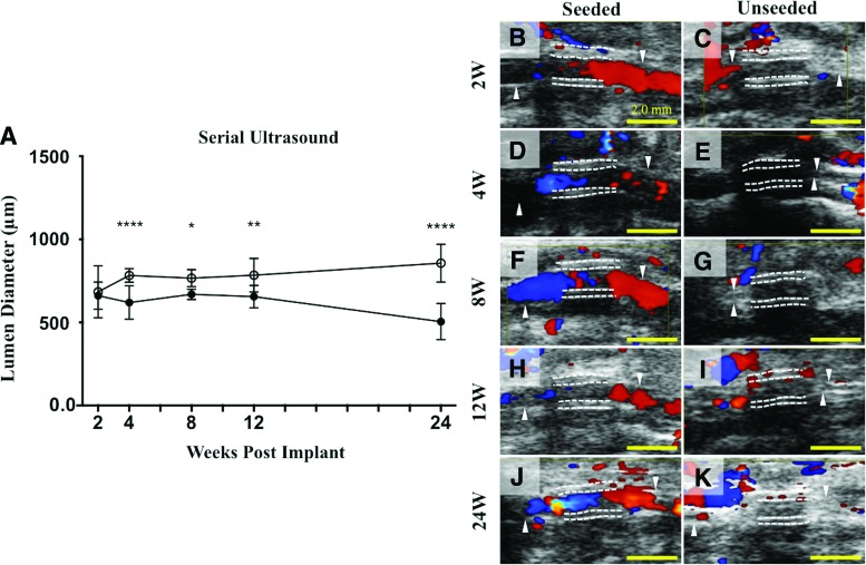
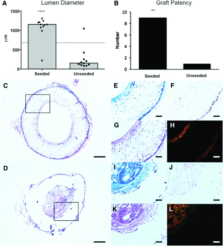
Methods: Nanofiber scaffolds were fabricated from co-electrospinning a solution of polyglycolic acid and a solution of poly(ι-lactide-co-ɛ-caprolactone) and characterized with scanning electron microscopy. Platelet activation and cell seeding efficiency were assessed by ATP secretion and DNA assays, respectively. Cell-free and BM-MNC seeded scaffolds were implanted in C57BL/6 mice (n = 15/group) as infrarenal inferior vena cava (IVC) interposition conduits. Animals were followed with serial ultrasonography for 6 months, after which grafts were harvested for evaluation of patency and neotissue formation by histology and immunohistochemistry (n = 10/group) and PCR (n = 5/group) analyses.

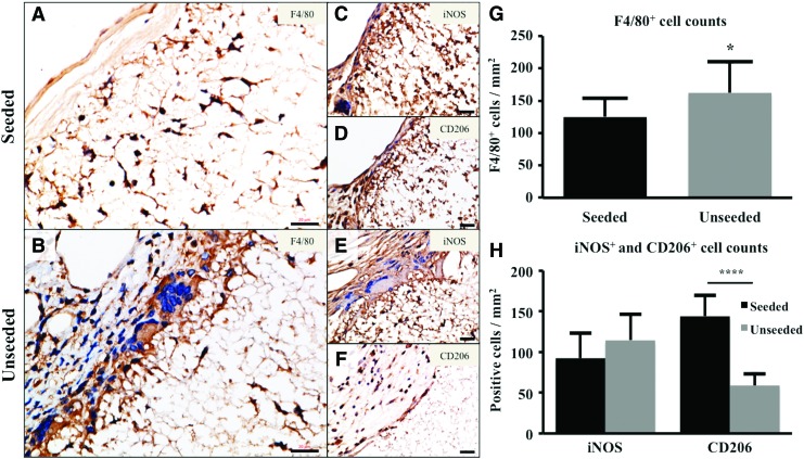
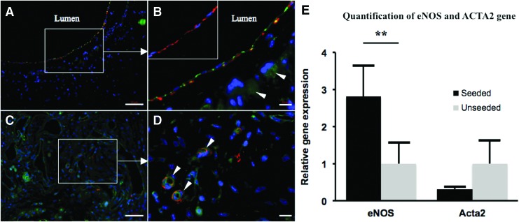
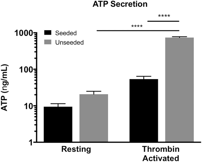
Results: BM-MNC seeding of electrospun scaffolds prevented stenosis compared with unseeded scaffolds (seeded: 9/10 patent vs. unseeded: 1/10 patent, p = 0.0003). Seeded vascular grafts demonstrated concentric laminated smooth muscle cells, a confluent endothelial monolayer, and a collagen-rich extracellular matrix. Platelet-derived ATP, a marker of platelet activation, was significantly reduced after incubating thrombin-activated platelets in the presence of seeded scaffolds compared with unseeded scaffolds (p < 0.0001). In addition, reduced macrophage infiltration and a higher M2 macrophage percentage were observed in seeded grafts.

Conclusions: The beneficial effects of BM-MNC seeding apply to electrospun TEVG scaffolds by attenuating stenosis through the regulation of platelet activation and inflammatory macrophage function, leading to well-organized neotissue formation. BM-MNC seeding is a valuable technique that can be used in the rational design of optimal TEVG scaffolds.

Keywords: biodegradable scaffold; bone marrow mononuclear cell (BM-MNC) seeding; electrospinning; nanofiber; stenosis; tissue-engineered vascular graft (TEVG).


Conflict of interest statement

Drs. Breuer and Shinoka receive research support from Gunze Ltd. (Kyoto, Japan) and Cook Regentec (Indianapolis, IN). Dr. Breuer is on the Scientific Advisory Board of Cook Medical (Bloomington, IN). Dr. Hibino receives research support from Secant Medical (Telford, PA). Jed Johnson is a co-founder of Nanofiber Solutions, Inc. (Columbus, OH). The remaining authors have no conflicts of interest to disclose.

Figures

<b>FIG. 1.</b>
FIG. 1.
Scaffold characterization. SEM images of luminal (A) and adventitial (B) surfaces of the electrospun scaffold. SEM of a BM-MNC-seeded scaffold luminal surface (C). Implantation of a seeded graft (D) in the murine infrarenal IVC interposition model. Fiber diameter (E) and pore size (F) for luminal surface (Fiber diameter: 0.61 ± 0.27 μm, pore size: 9.71 ± 0.16 μm). In vitro degradation mechanical values of UTS (G), percent elongation to failure (H), and Young's Modulus (I). Scale bar = 50 μm. BM-MNC, bone marrow mononuclear cell; IVC, inferior vena cava; UTS, ultimate tensile strength. Color images available online at www.liebertpub.com/tea
<b>FIG. 2.</b>
FIG. 2.
Lumen diameter over time by serial ultrasound. Serial ultrasound of implanted TEVGs performed at 2, 4, 8, 12, and 24 weeks after graft implantation indicated a significant difference in lumen diameter between the seeded and unseeded groups as early as 4 weeks after implantation that was maintained throughout the 6-month time course (A), ****p < 0.0001, *p < 0.05, **p < 0.005. Representative ultrasound images from each time point (B–K), respectively, demonstrating patent grafts characterized by color Doppler flow through the lumen (B, D, F, H, J) in the seeded group and occluded grafts in the unseeded group (C, E, G, I, K). Color Doppler signals in the unseeded group may indicate the presence of collateral vasculature surrounding stenotic TEVGs. The white dotted lines indicate the luminal and adventitial surfaces of the TEVGs. Scale bar = 2.0 mm. TEVG, tissue-engineered vascular graft. Color images available online at www.liebertpub.com/tea
<b>FIG. 3.</b>
FIG. 3.
Histological assessment of vascular neotissue formation after 6 months. Histomorphometric comparison of the lumen diameter measurements (A) and graft patency (B) between the seeded and unseeded groups demonstrates that BM-MNC seeding prevents occlusion in the nanofiber scaffold (****p < 0.0001, ***p = 0.0003). Histologic comparison between seeded and unseeded scaffolds demonstrated vascular neotissue formation in seeded grafts characterized by mature ECM on the luminal and adventitial graft surfaces, adequate cellular infiltration, and minimal vascular calcification. Representative photomicrographs for each group (Seeded: [C, E–H]; Unseeded: D, I–L) are shown for H&E (C, D), Masson's trichrome (E, I), Hart's (G, K), von Kossa (F, J), and Picrosirius red stains (H, L). Scale bar = 200 μm for (C) and (D), and 100 μm otherwise. ECM, extracellular matrix. Color images available online at www.liebertpub.com/tea
<b>FIG. 4.</b>
FIG. 4.
BM-MNC seeding of electrospun TEVG modulated host inflammatory cell infiltration and phenotype. F4/80 staining of seeded (A) and unseeded (B) electrospun TEVGs. Phenotypic characterization of TEVG macrophages demonstrated iNOS+ (Seeded: C; Unseeded: E) and CD206+ (Seeded: [D]; Unseeded: [F]) cells. F4/80+ cells in the seeded vs. unseeded group at 6 months (G); seeded group revealed significantly less macrophage infiltration (*p < 0.05). iNOS+ and CD206+ cells in the seeded versus unseeded group at 6 months (H); unseeded group showed significantly less CD206+ cells (****p < 0.0001). Scale bar = 20 μm. Color images available online at www.liebertpub.com/tea
<b>FIG. 5.</b>
FIG. 5.
Cell-seeded nanofiber scaffolds are characterized by well-organized and mature vascular neotissue. Immunofluorescent staining of BM-MNC-seeded scaffolds indicated the formation of well-organized neo-intima characterized by a medial α-SMA+ (red) smooth muscle cell layer lined by CD31+ (green) endothelial cells (A, B, arrowheads denote background staining due to remaining scaffold material). SM-MHC staining (red) confirmed that the medial smooth muscle cells in the seeded group were mature and contractile (B, inset). Unseeded TEVG neotissue was characterized by an unorganized population of α-SMA+ and CD-31+ cells (C), with evidence of re-canalization (D, arrowheads indicate neovascularization of stenotic tissue). Quantification of eNOS and ACTA2 gene transcription by RT-qPCR (E); less ACTA2 and significantly more eNOS gene transcription in the seeded group (**p < 0.005). Scale bar = 50 μm for (A, C), 10 μm for (B, D). RT-qPCR, reverse transcription-quantitative polymerase chain reaction. Color images available online at www.liebertpub.com/tea
<b>FIG. 6.</b>
FIG. 6.
Cell seeding reduced measurable ATP from thrombin-activated platelets. The quantity of ATP derived from thrombin-activated platelets in the presence of BM-MNC-seeded scaffolds was significantly less than in the presence of unseeded scaffolds (****p < 0.001). In addition, after incubation with unseeded scaffolds, a significant difference in the amount of ATP derived from thrombin-activated and resting platelets was found (****p < 0.001), but no difference was observed in the seeded group, suggesting that BM-MNC seeding may regulate platelet function.

Similar articles

Cited by

References

    1. Pham Q.P., Sharma U., and Mikos A.G. Electrospinning of polymeric nanofibers for tissue engineering applications: a review. Tissue Eng 12, 1197, 2006 - PubMed
    1. Cleary M.A., Geiger E., Grady C., Best C., Naito Y., and Breuer C. Vascular tissue engineering: the next generation. Trends Mol Med 18, 394, 2012 - PubMed
    1. Rocco K.A., Maxfield M.W., Best C.A., Dean E.W., and Breuer C.K. In vivo applications of electrospun tissue-engineered vascular grafts: a review. Tissue Eng Part B Rev 20, 628, 2014 - PubMed
    1. Tara S., Rocco K.A., Hibino N., et al. . Vessel bioengineering. Circ J 78, 12, 2014 - PubMed
    1. Rathore A., Cleary M., Naito Y., Rocco K., and Breuer C. Development of tissue engineered vascular grafts and application of nanomedicine. Wiley Interdiscip Rev Nanomed Nanobiotechnol 4, 257, 2012 - PubMed

Publication types