. 2017 May 9;12(5):e0177224.
doi: 10.1371/journal.pone.0177224. eCollection 2017.

Strong upregulation of inflammatory genes accompanies photoreceptor demise in canine models of retinal degeneration

Affiliations

Strong upregulation of inflammatory genes accompanies photoreceptor demise in canine models of retinal degeneration

Tatyana Appelbaum et al. PLoS One. .

Abstract

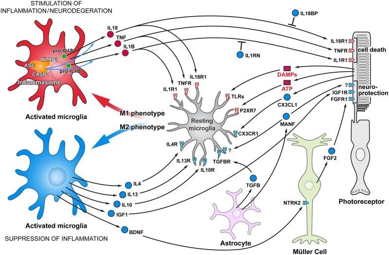
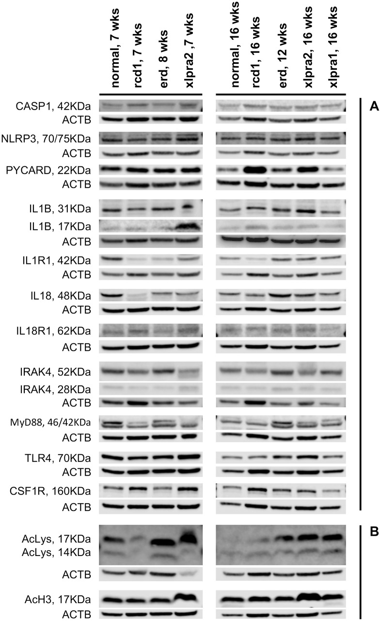
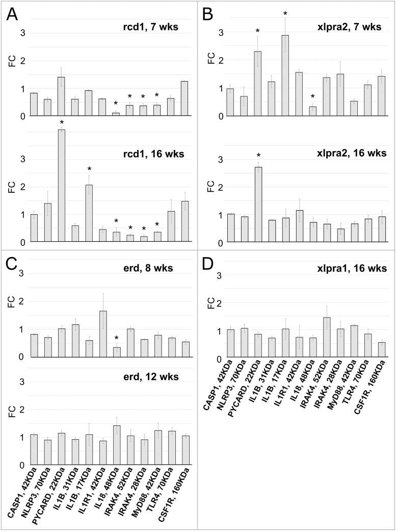
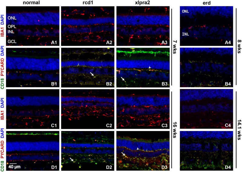
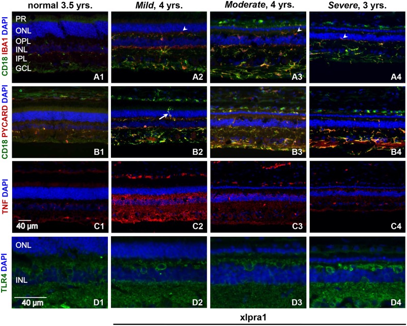
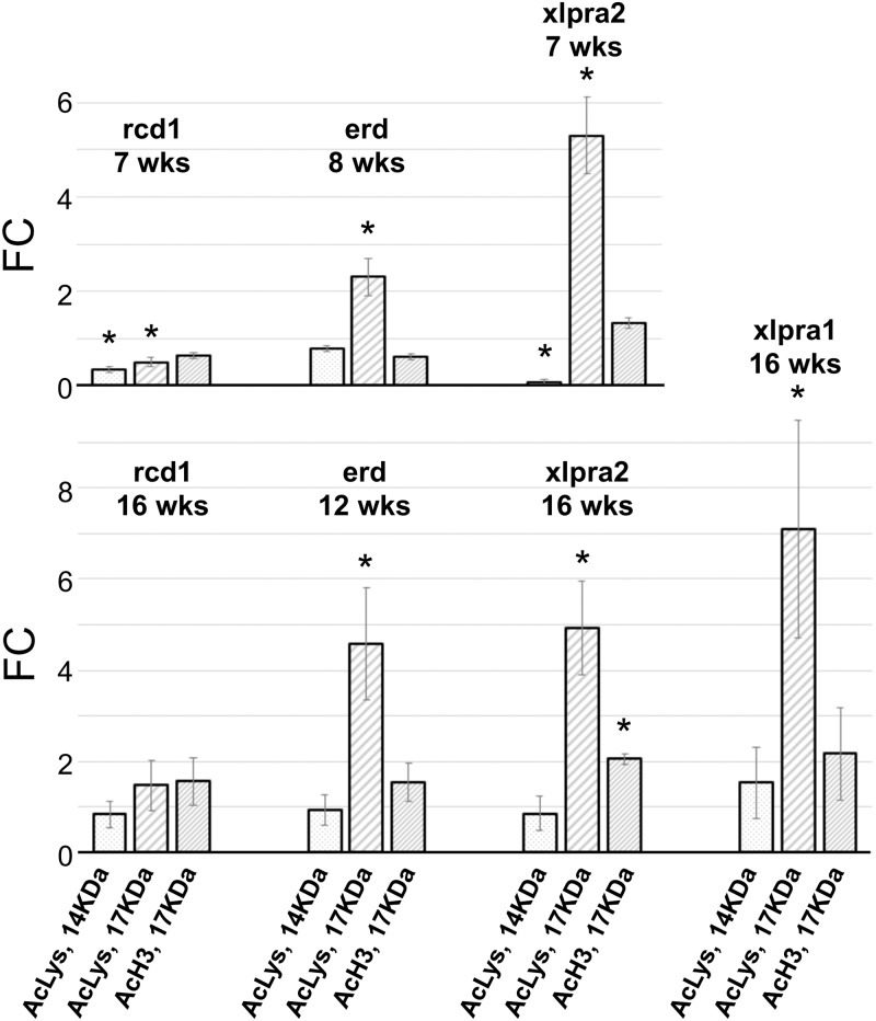
We have analyzed the complex pattern of the inflammatory response in early-onset canine models of human retinitis pigmentosa, rcd1, xlpra2 and erd, as well as late-onset xlpra1, in comparative manner. The time course of immune response genes and proteins expression was examined along the timeline of photoreceptors degeneration. Gene expression analysis of the early-onset models prior to and after the peak of photoreceptors death identified the involvement of multiple immune response genes including those encoding constituents of the NLRP3 inflammasome, its substrates, pro-IL1B, pro-IL18, and common components of IL1B, IL18 and TLR4 pathways. Out of two activated caspase-1 cleavage products, IL1B and IL18, only IL1B was detected in rcd1 and xlpra2 while precursor IL18 remained unprocessed in the same protein extract highlighting prominence of IL1B pathway. An overall immune response was most prominent in rcd1 followed by xlpra2 and least prominent in erd. Noticeably, in rcd1 and xlpra2, but not in erd, early induction of the immune response was accompanied by sustained intraretinal migration and activation of retinal microglia. Lastly, delayed activation of the anti-inflammatory factors in all early-onset models was insufficient to counterbalance rapidly progressing inflammation. In contrast to early-onset models, in late-onset xlpra1 retinas a subset of the pro-inflammatory genes was highly upregulated long before any disease-related structural changes occurred, but was counterbalanced by an adequate anti-inflammatory response. Results point out to upregulated immune response accompanying disease progression in animal models of retinal degeneration, and to potential benefits of early anti-inflammatory therapy.


Conflict of interest statement

Competing Interests: The authors have declared that no competing interests exist.

Figures

Fig 1
Fig 1. Functional cell-cell interactions in retina responsible for stimulating and suppressing inflammation.
Schematic representation of glia-photoreceptor interactions in the retina. The connections are based on reported cellular interactions (for description, please see text). Microglia, the resident immune cells, are responsible for initiating an inflammatory response. Microglia activation in the degenerating retina is triggered by different DAMPs. Although some trigger molecules which activate microglia are predicted to be released from injured cells, e.g. ATP, other molecules have not yet been identified. Activated microglia are capable of acquiring diverse phenotypes that display different cell-surface and intracellular markers, secrete different factors, and exhibit different functions. Two extreme microglial phenotypes are shown: the classically activated (M1) phenotype that promotes a pro-inflammatory response, and the alternatively activated (M2) phenotype that facilitates an anti-inflammatory response. Microglia can control their own polarization through autocrine and paracrine mechanisms. M1-polarized microglial phenotype is promoted by several cytokines, including TNF, IL1B and IL18. On the other hand, microglia can be driven to M2 phenotype by stimuli like IL4, IL13, IL-10, TGFB, CX3CL1 and MANF. Microglia, using receptors and signals, are in constant communication with neurons and other retinal cells. In pathological conditions this tight communication between cells mediate adaptive responses within the retina. Comments: Three basic types of glial cell in the retina are shown: Müller cells, astroglia, and microglia phenotypes are resting or activated, M1 or M2. Pro-inflammatory cytokines/receptors are in red and anti-inflammatory/neuroprotective factors are marked in blue. In addition, since MANF receptor is not identified yet, it’s marked by “?”.
Fig 2
Fig 2. Western blot analysis of pro-inflammatory (A) and histone acetylation proteins (B) in study models of retinal degeneration.
(A) Representative western immunoblot was performed for the expression of 11 proteins involved in pro-inflammatory signaling in total retinal protein extracts for normal and mutant retinas at 7 wks (rcd1, xlpra2), 8 wks (erd), 12 wks (erd), and 16 wks (rcd1, xlpra2, xlpra1). The following proteins were analyzed: inflammasome components (CASP1, NLRP3 and PYCARD), inflammasome substrates (IL1B and IL18) and their receptors (IL1R1 and IL18R1), inflammasome receptor (TLR4), common components of IL1B-, IL18- and TLR4-pathways (MYD88 and IRAK4) and macrophages expressing protein (CSF1R). (B) Level of histone acetylation in retinal protein extracts from the same four disease models was evaluated with acetylated-Lysine and acetyl-Histone H3 antibodies at the indicated time points.
Fig 3
Fig 3. Protein quantification in retinas of rcd1 (A), xlpra2 (B), erd (C) and xlpra1 (D) models.
Quantification of pro-inflammatory proteins on western blots (Fig 2A) normalized against the corresponding housekeeping protein (ACTB) was done using Li-COR Odyssey Fc and represented as fold-changes compared to the normal tissue values. Differences in relative fluorescence (Y-axis) for the proteins analyzed (X-axis) show CASP1, NLRP3, IL1B-precursor, IL1R1, TLR4 and CSF1R in disease (rcd1, xlpra2, erd, xlpra1) were similar to normal levels at all ages. In contrast, two proteins were increased over normal levels: mature ILIB-17 KDa (7 wks xlpra2, 16 wks rcd1) and PYCARD (7 wks xlpra2, 16 wks xlpra2, 16 wks rcd1). Moreover, three proteins were below normal levels: MYD88 (7 wks rcd1, 16 wks rcd1), IRAK4 (7 wks rcd1, 16 wks rcd1) and IL18-precursor (7 wks rcd1, 16 wks rcd1, 8 wks erd, 7 wks xlpra2). * indicates significance level 5% (p<0.05).
Fig 4
Fig 4. Retinal localization of selected pro-inflammatory proteins in early-onset disease models.
Immunolabeling of normal and early-onset disease (rcd1, erd, xlpra2) retinas at various ages using IBA1, PYCARD and CD18 antibodies. IBA1 (red) labeling, a well-known marker of reactive microglia, increases in rcd1 and xlpra2 at 7 and 16 wks (A2, A3, C2, C3) when compared to normal control (A1, C1), suggesting microglial activation. IBA+ cells demonstrate migration of microglia from inner retina towards the outer retinal layers of rcd1 (A2, C2) and xlpra2 (A3, C3) at all ages tested. Additionally, double immunolabeling of the same retinal samples were also done with microglia/macrophage marker CD18 (green) and the inflammasome component PYCARD (red) antibodies. PYCARD is expressed primarily in CD18+ positive cells, as shown by labeling overlapping (B2-B4, D2-D4) thus demonstrating inflammasome component expression specifically in retinal microglia, albeit in two distinct subpopulations; CD18+/PYCARD+ (yellow; arrow) and CD18-/PYCARD+ (red; arrowhead) cells. PYCARD expression is observed at all studied ages in diseased (B2-B4, D2-D4) and normal (B1, D1) retinas, and is significantly upregulated in rcd1 and xlpra2, especially at 16 wks of age. Conversely, IBA1 staining in erd retinas at 8 wks (A4) and 14.1 wks (C4) do not significantly change when compared to normal (A1, C1) indicating no additional microglial activation is occurring. Similarly, PYCARD/CD18 staining in erd at 8 and 14.1 wks remains the same as normal retinas. Although two subpopulations of CD18/PYCARD cells are also observed in erd retinas, increase in microglia migration to the outer retina layers as seen in rcd1 and xlpra2 was noticeably absent. ONL = outer nuclear layer; OPL = outer plexiform layer; INL = inner nuclear layer; GCL = ganglion cells layer. Scale bar 40 μm.
Fig 5
Fig 5. Retinal localization of pro-inflammatory proteins in late-onset xlpra1-affected retinas with different disease severity phenotypes.
Immunolabeling of normal and late-onset disease (xlpra1) retinas with Mild, Moderate and Severe phenotypes was done using IBA1, CD18, PYCARD, TNF and TLR4 antibodies. Double immunolabeling with microglial markers CD18 (green) and IBA1 (red) antibodies was done (A1–A4) to describe the phagocytes response characterized by their morphological changes and intraretinal migration. IBA1 and CD18 show significant co-localization in xlpra1 retinas displaying a well-developed disease phenotype that includes reduction of ONL (A2-A4). Both IBA1 and CD18 immunolabeling are robustly upregulated in disease retinas although highest expression is seen in Moderate severity. Double staining (A1-A4) of IBA1 and CD18 shows three subpopulation of phagocytes are seen in xlpra1 retina: CD18+/IBA1+ (yellow), CD18+/IBA1- (green) and CD18-/IBA1+ (red). Additionally, aggressive invasion of retinal layers by activated microglia/macrophages (arrowhead) is noticeable (A2-A4), particularly at Mild stage. Double immunolabeling of CD18 (green) with PYCARD (red) antibodies (B1-B4) shows PYCARD expression in CD18+ positive cells (yellow), with upregulation of both proteins in affected retinas. Robust infiltration of ONL by activated microglia/macrophages (B2) is very prominent in Mild stage (arrow). TNF labeling in normal and diseased retinas (C1-C4) shows TNF present in all retinal layers following the same pattern at all disease stages analyzed, however in Mild disease labeling signal was highest while intensity decreases as disease progresses. TLR4 labeling is present in all layers of normal and diseased retinas with labeling higher in Severe phenotype. PR = photoreceptors; ONL = outer nuclear layer; OPL = outer plexiform layer; INL = inner nuclear layer; IPL = inner plexiform layer; GCL = ganglion cells layer. Scale bar 40 μm.
Fig 6
Fig 6. Quantification of histone acetylation level in rcd1, xlpra2, erd and xlpra1 retinas.
Quantification of histone acetylation on western blots (Fig 2B) normalized against the corresponding housekeeping protein (ACTB) was done using Li-COR Odyssey Fc and represented as fold-changes compared to the normal tissue values. Differences in relative fluorescence (Y-axis) for the proteins analyzed (X-axis) show 14 KDa-Acetyl-Lysine levels decrease in rcd1 and xlpra2 at 7 wks but remain the same as normal in all other samples. 17 KDa-Acetyl-Lysine was increased in all samples except in 7 wks rcd1 (decreased) and 16 wks rcd1 (same as normal). In contrast, acetylated H3 histone shows mostly unchanged pattern, except in 16 wks xlpra2 where it was increased. * indicates significance level 5% (p<0.05).

Similar articles

Cited by

References

    1. Eandi CM, Charles Messance H, Augustin S, Dominguez E, Lavalette S, Forster V, et al. Subretinal mononuclear phagocytes induce cone segment loss via IL-1beta. Elife. 2016;5: - PMC - PubMed
    1. Gupta N, Brown KE, Milam AH. Activated microglia in human retinitis pigmentosa, late-onset retinal degeneration, and age-related macular degeneration. Exp Eye Res. 2003;76: 463–471. - PubMed
    1. Whitcup SM, Nussenblatt RB, Lightman SL, Hollander DA. Inflammation in retinal disease. Int J Inflam. 2013;2013: 724648 10.1155/2013/724648 - DOI - PMC - PubMed
    1. Yoshida N, Ikeda Y, Notomi S, Ishikawa K, Murakami Y, Hisatomi T, et al. Clinical evidence of sustained chronic inflammatory reaction in retinitis pigmentosa. Ophthalmology. 2013;120: 100–105. 10.1016/j.ophtha.2012.07.006 - DOI - PubMed
    1. Rock KL, Kono H. The inflammatory response to cell death. Annu Rev Pathol. 2008;3: 99–126. 10.1146/annurev.pathmechdis.3.121806.151456 - DOI - PMC - PubMed

Substances

LinkOut - more resources