. 2017 May 10;14(1):103.
doi: 10.1186/s12974-017-0874-x.

A role for cathepsin Z in neuroinflammation provides mechanistic support for an epigenetic risk factor in multiple sclerosis

Affiliations

A role for cathepsin Z in neuroinflammation provides mechanistic support for an epigenetic risk factor in multiple sclerosis

Euan R O Allan et al. J Neuroinflammation. .

Abstract

Background: Hypomethylation of the cathepsin Z locus has been proposed as an epigenetic risk factor for multiple sclerosis (MS). Cathepsin Z is a unique lysosomal cysteine cathepsin expressed primarily by antigen presenting cells. While cathepsin Z expression has been associated with neuroinflammatory disorders, a role for cathepsin Z in mediating neuroinflammation has not been previously established.

Methods: Experimental autoimmune encephalomyelitis (EAE) was induced in both wildtype mice and mice deficient in cathepsin Z. The effects of cathepsin Z-deficiency on the processing and presentation of the autoantigen myelin oligodendrocyte glycoprotein, and on the production of IL-1β and IL-18 were determined in vitro from cells derived from wildtype and cathepsin Z-deficient mice. The effects of cathepsin Z-deficiency on CD4+ T cell activation, migration, and infiltration to the CNS were determined in vivo. Statistical analyses of parametric data were performed by one-way ANOVA followed by Tukey post-hoc tests, or by an unpaired Student's t test. EAE clinical scoring was analyzed using the Mann-Whitney U test.

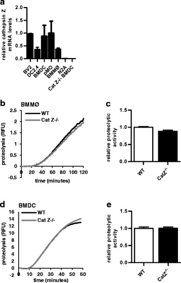
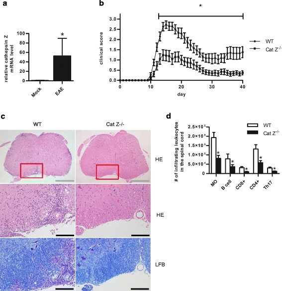
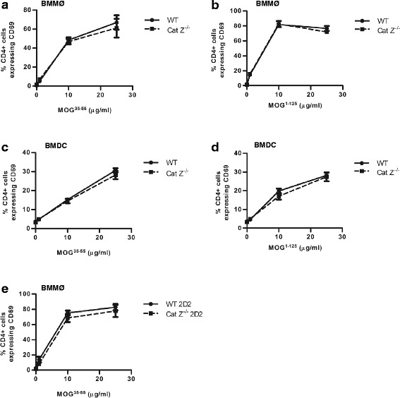
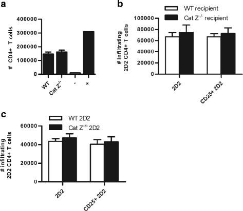
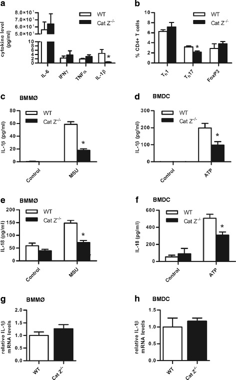
Results: We showed that mice deficient in cathepsin Z have reduced neuroinflammation and dramatically lowered circulating levels of IL-1β during EAE. Deficiency in cathepsin Z did not impact either the processing or the presentation of MOG, or MOG- specific CD4+ T cell activation and trafficking. Consistently, we found that cathepsin Z-deficiency reduced the efficiency of antigen presenting cells to secrete IL-1β, which in turn reduced the ability of mice to generate Th17 responses-critical steps in the pathogenesis of EAE and MS.

Conclusion: Together, these data support a novel role for cathepsin Z in the propagation of IL-1β-driven neuroinflammation.


Figures

Fig. 1
Fig. 1
Cathepsin Z is highly expressed in APCs but does not significantly contribute to phagolysosomal proteolysis. a Cathepsin Z mRNA levels in BV2 (C57BL/6 brain microglia cell line), DC2.4 (dendritic cell line), BMDC (bone marrow derived dendritic cells), pMØs (peritoneal macrophages), BMMØ (bone marrow derived macrophages), N2A (murine albino neuroblastoma cell line) and Cat Z−/− BMDCs (n = 3). b-e The total proteolytic activity (rate of substrate-liberated fluorescence from the particle-bound fluorogenic substrate DQ-albumin) following phagocytosis of fluorometric experimental particles in WT and Cat Z−/− (b-c) BMMØ (n = 9) and (d-e) BMDC (n = 5). b, d Representative real-time traces of phagosomal proteolysis. c, e Averaged rates of proteolysis (determined by calculation of the slope of the linear portion of the real-time trace [as described by y = mx + c, where y = relative fluorescence, m = slope, and x = time] were calculated between (c) 20 min and 60 min or (e) 20 min and 40 min after particle internalization. Data presented as mean+/− SEM; (c, e) no significant differences (unpaired Student’s t-test, p > 0.05) from the WT control were observed
Fig. 2
Fig. 2
Cathepsin Z expression is increased in the CNS during EAE, and mice deficient in cathepsin Z exhibit attenuated signs of neuroinflammation and demyelination during EAE. a Cathepsin Z mRNA levels in the spinal cord tissue of WT mice 15 days after induction of EAE or mock (n = 6). b Clinical disease course of WT and Cat Z−/− mice after active induction of EAE (n = 20–21). c Representative micrographs of transverse sections of lumbar spinal cord from WT and Cat Z−/− mice at 15 days after induction of EAE. Sections are stained with hematoxylin and eosin (HE) for inflammation, or Luxol fast blue (LFB) for demyelination. Grey and black scale bars indicate 500 and 100 μm respectively. d The total number of infiltrating macrophages (MO), B cells, CD8+ T cells (CD8+), CD4+ T cells (CD4+), and Th17 cells (IL-17+/CD4+) isolated from lumbar spinal cord tissue 15 days post EAE induction as analyzed by flow cytometry (n = 8–12). Data presented as mean +/− SEM; significant differences (unpaired Student’s t test; clinical data, Mann–Whitney U test; p < 0.05) from the WT control are denoted by asterisks
Fig. 3
Fig. 3
Cathepsin Z-deficiency does not affect the efficiency of MOG antigen processing or presentation in BMMØ and BMDC, or the efficiency of MOG-specific CD4+ T cell activation. a-e WT and Cat Z−/− (a, b, e) BMMØ and (c-d) BMDC were incubated with (a, c, e) MOG35–55 peptide (0, 1, 10, 25 μg/ml), or (b, d) recombinant MOG1–125 (0, 1, 10, 25 μg/ml) for 6 h before co-incubation with (a-d) MOG35–55-specific 2D2 CD4+ T cells or (e) WT or Cat Z−/− MOG35–55-specific 2D2 CD4+ T cells. Activation of WT and Cat Z−/− MOG35–55-specific 2D2 CD4+ T cells was assessed via CD4+ T cell CD69 surface expression after 16 h co-incubation with APCs (n = 3–6). Data presented as mean+/− SEM; no significant differences (unpaired Student’s t test, p > 0.05) from WT controls were observed
Fig. 4
Fig. 4
CD4+ T cells deficient in cathepsin Z exhibit proficient migration in vitro; and efficient trafficking, infiltration, and reactivation in the CNS. a To determine whether deficiency of cathepsin Z affected the ability of CD4+ T cells to undergo chemotaxis in response to CXCL9, CD4+ T cells were isolated from the spleens of WT and Cat Z−/− mice and given 1 h to migrate through an ICAM coated Transwell plate in response to CXCL9 (n = 4). The negative control (−) well had no CXCL9. The positive control (+) well had no filter, allowing all CD4+ T cells to migrate through to the bottom of the Transwell. b To evaluate the ability of CD4+ T cells to infiltrate the CNS in WT and Cat Z−/− mice, MOG35–55-specific 2D2 CD4+ T cells were isolated and expanded ex vivo using IL-12 and MOG35–55 for 48 h before adoptive transfer into WT and Cat Z−/− recipient mice; alternatively, (c) to examine the capacity of Cat Z−/− CD4+ T cells to infiltrate the CNS of WT mice, WT and Cat Z−/− MOG35–55-specific 2D2 CD4+ T cells were isolated, expanded and adoptively transferred into WT recipients. b-c Six days following adoptive transfer, the 2D2 CD4+ T cells were isolated from the CNS using a discontinuous Percoll gradient, identified by flow cytometry (CD4+, Vα3.2+) and evaluated for the expression of the activation marker CD25 (n = 4). Data presented as mean+/− SEM; no significant differences (unpaired Student’s t test, p > 0.05) from the WT control were observed
Fig. 5
Fig. 5
Mice deficient in cathepsin Z have dramatically reduced circulating IL-1β during EAE and attenuated Th17 responses in vivo; consistently, APCs deficient in cathepsin Z are unable to efficiently generate IL-1β and IL-18 in vitro. a Proinflammatory cytokine levels in WT and Cat Z−/− EAE serum 15 days post induction (n = 3–6). b The percentage of Th1 (IFNγ+), Th17 (IL-17+), and FoxP3+ CD4+ T cells isolated from the inguinal lymph nodes of WT and Cat Z−/− mice 6 days after injection with CFA (n = 3). c, d Concentration of IL-1β within the supernatant of WT and Cat Z−/− (c) BMMØ, and (d) BMDC after priming with LPS and subsequent exposure to NLRP3-inflammasome activators (c) monosodium urate (MSU) or (d) ATP as quantified by ELISA (n = 5). e, f Concentration of IL-18 within the supernatant of WT and Cat Z−/− (e) BMMØ, and (f) BMDC after priming with LPS and subsequent exposure to the NLRP3-inflammasome activators (e) MSU or (f) ATP as quantified by ELISA (n = 5). g, h IL-1β mRNA levels of WT and Cat Z−/− (g) BMMØ and (h) BMDCs stimulated with LPS as determined by qPCR (n = 3–5). Data presented as mean +/− SEM; significant differences (unpaired Student’s t test, p < 0.05) from the WT control are denoted by asterisks

References

    1. Consortium IMSG, 2 WTCCC Genetic risk and a primary role for cell-mediated immune mechanisms in multiple sclerosis. Nature. 2011;476:214–9. doi: 10.1038/nature10251. - DOI - PMC - PubMed
    1. Huynh JL, Garg P, Thin TH, Yoo S, Dutta R, Trapp BD, Haroutunian V, Zhu J, Donovan MJ, Sharp AJ. Epigenome-wide differences in pathology-free regions of multiple sclerosis-affected brains. Nat Neurosci. 2014;17:121–30. doi: 10.1038/nn.3588. - DOI - PMC - PubMed
    1. Nägler DK, Ménard R. Human cathepsin X: a novel cysteine protease of the papain family with a very short proregion and unique insertions. FEBS Lett. 1998;434:135–9. doi: 10.1016/S0014-5793(98)00964-8. - DOI - PubMed
    1. Santamaria I, Velasco G, Pendas AM, Fueyo A, Lopez-Otin C. Cathepsin Z, a novel human cysteine proteinase with a short propeptide domain and a unique chromosomal location. J Biol Chem. 1998;273:16816–23. doi: 10.1074/jbc.273.27.16816. - DOI - PubMed
    1. Nägler DK, Zhang R, Tam W, Sulea T, Purisima EO, Ménard R. Human cathepsin X: A cysteine protease with unique carboxypeptidase activity. Biochemistry. 1999;38:12648–54. doi: 10.1021/bi991371z. - DOI - PubMed

Publication types

MeSH terms