

Small, smooth, nonmobile cardiac myxoma detected by transesophageal echocardiography following recurrent cerebral infarction: a case report
- PMID: 28486992
- PMCID: PMC5424306
- DOI: 10.1186/s13256-017-1298-z
Small, smooth, nonmobile cardiac myxoma detected by transesophageal echocardiography following recurrent cerebral infarction: a case report
Abstract
Background: Cardiac myxoma is known to cause repeated events of cerebral embolism. Soft and irregularly shaped myxomas with high mobility are associated with a higher occurrence of cerebral embolism. In contrast, nonmobile cardiac myxomas with a round regular shape are rarely considered to be a cause of cerebral embolism. In this case, we present a patient with recurrent cerebral embolism associated with a small and nonmobile cardiac myxoma of round regular shape.
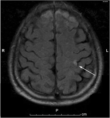
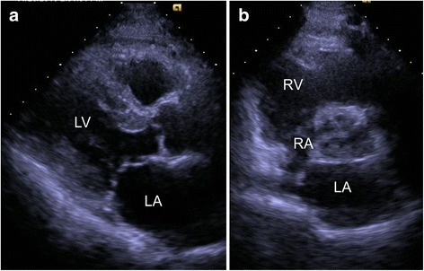
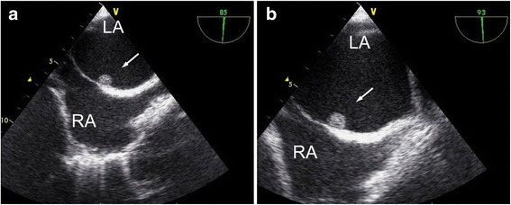
Case presentation: A 76-year-old Japanese man presented to our hospital with weakness in his right upper extremity. He had a history of right frontal lobe infarction in the previous month. T2-weighted magnetic resonance imaging revealed an area of hyperintensity in the left precentral gyrus, indicating acute cerebral infarction. Transthoracic echocardiography revealed normal left ventricular function and no abnormalities. However, transesophageal echocardiography showed a small and nonmobile left atrial tumor with round regular shape attached to the ostium secundum of the atrial septum. Based on these findings, we diagnosed recurrent cerebral infarction due to embolization caused by left atrial myxoma, and cardiac tumor extraction was performed on hospitalization day 36. The excised tumor measured 0.6 × 0.6 × 0.5 cm and was diagnosed as cardiac myxoma by histologic examination.
Conclusions: Even small and nonmobile cardiac myxomas with a round regular shape may cause recurrent cerebral infarction. The diagnosis of this type of atrial myxoma is elusive and transesophageal echocardiography was an effective method of detection. In a clinical situation, this type of cardiac myxoma may be overlooked as a cause of cerebral infarction.
Keywords: Cardiac tumor; Cerebral infarction; Echocardiography.
Figures

References
Publication types
MeSH terms
LinkOut - more resources
Full Text Sources
Other Literature Sources
