Review
. 2017 Sep;22(9):1336-1350.
doi: 10.1016/j.drudis.2017.04.021. Epub 2017 May 6.

Development of nanomaterials for bone-targeted drug delivery

Affiliations
Review

Development of nanomaterials for bone-targeted drug delivery

Hao Cheng et al. Drug Discov Today. 2017 Sep.

Abstract

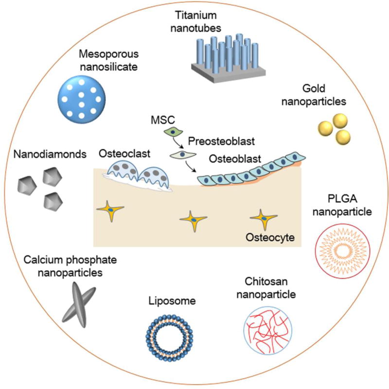
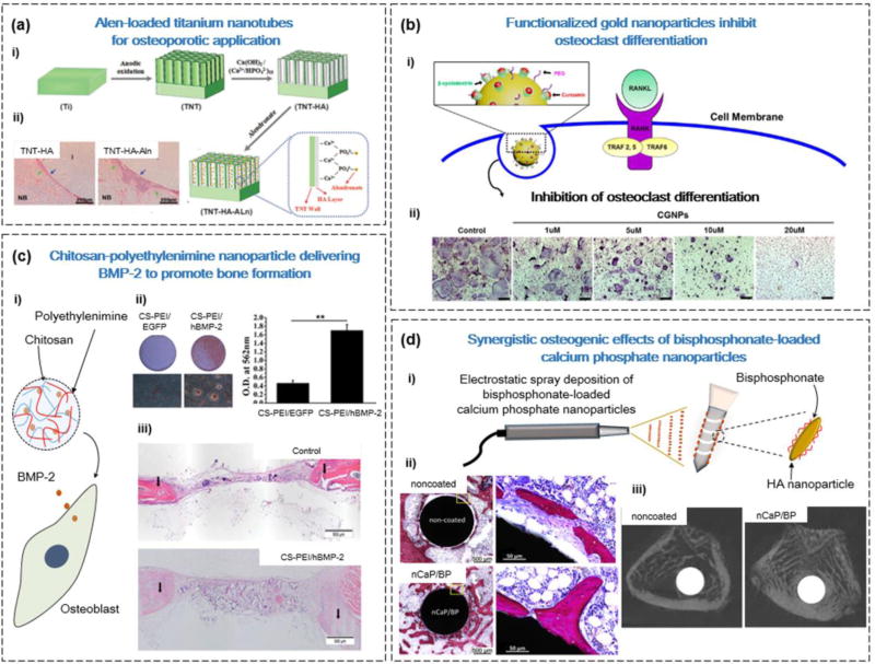
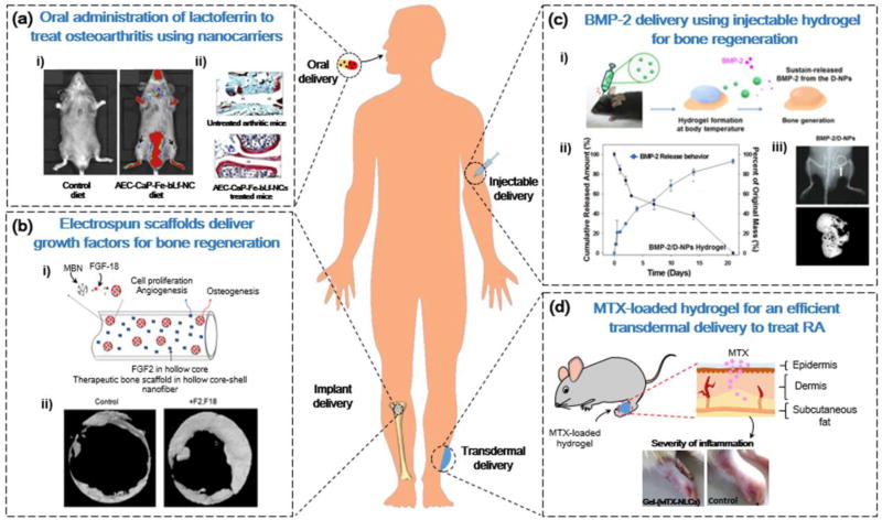
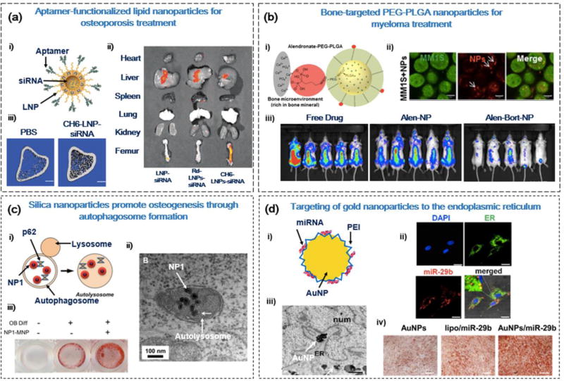
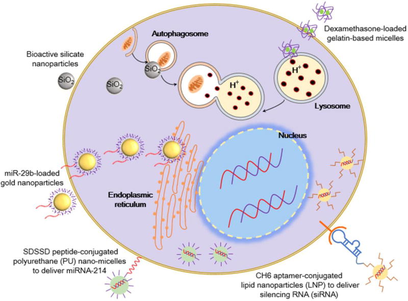
Bone is one of the major organs of the human body; it supports and protects other organs, produces blood cells, stores minerals, and regulates hormones. Therefore, disorders in bone can cause serious morbidity, complications, or mortality of patients. However, despite the significant occurrence of bone diseases, such as osteoarthritis (OA), osteoporosis (OP), non-union bone defects, bone cancer, and myeloma-related bone disease, their effective treatments remain a challenge. In this review, we highlight recent progress in the development of nanotechnology-based drug delivery for bone treatment, based on its improved delivery efficiency and safety. We summarize the most commonly used nanomaterials for bone drug delivery. We then discuss the targeting strategies of these nanomaterials to the diseased sites of bone tissue. We also highlight nanotechnology-based drug delivery to bone cells and subcellular organelles. We envision that nanotechnology-based drug delivery will serve as a powerful tool for developing treatments for currently incurable bone diseases.


Figures

Figure 1
Figure 1
Examples of nanomaterials for bone drug delivery. The inorganic nanomaterials include titanium nanotubes, gold nanoparticles, calcium phosphate nanoparticles, and mesoporous silica nanoparticles. The organic nanomaterials include chitosan nanoparticles, poly(L-lactide-co-glycolide) (PLGA) nanoparticles, and liposomes. These nanomaterials can selectively target bone tissues and cells to deliver drugs. Abbreviation: MSC, mesenchymal stem cell.
Figure 2
Figure 2
Types of nanomaterial for applications in bone drug delivery. (a) Alendronate (Alen)-loaded titanium nanotubes (TNTs) were able to promote bone formation in a rabbit osteoporosis model. (i) Fabrication of hydroxyapatite (HA)-coated TNTs for local delivery of Alen. (ii) Hemotoxylin and eosin (H&E) images of TNT-HA and TNT-HA-Alen, showing more newly formed bone in the TNT-HA-Alen group (blue arrows). (b) Delivery of the drug curcumin using βCD-functionalized gold nanoparticles (GNPs) to inhibit osteoclast differentiation. (i) Schematic of the GNPs with βCD-curcumin inclusion complexes for intracellular delivery of the drug. (ii) Dose-dependent reduction of TRAP expression with administration of GNPs functionalized with βCD-curcumin. (c) Delivery of the gene encoding bone morphogenetic protein 2 (BMP2) using chitosan (CS)-polyethylenimine (PEI) nanoparticles to promote bone formation. (i) CS-PEI nanoparticles can efficiently transfect osteoblasts with the BMP2 gene. (ii) CS-PEI/BMP-2 promotes mineralization, determined by Alizarin red staining 21 days after transfection. Mineralization was quantified by adding cetylpyridinium chloride. (iii) Implantation of the CS-PEI/BMP-2 collagen scaffolds promoted new bone formation, determined by H&E staining. (d) The coating of bone implants with calcium phosphate (CaP) nanoparticles (CPNs) and bisphosphonates (BPs) can improve their integration with bone tissue. (i) Using the electrospray deposition technique, the surface of bone implants can be functionalized with CPNs and BP. (ii) Histological sections of bone tissue demonstrated that CPN and/or BP functionalized implants form direct contact with bone tissue. Scale bars = 500 Mm (left) and 50 Mm (right). (iii) Micro-CT images of bone tissue after 4 weeks with noncoated and coated bone implants demonstrated improved bone volume percentage. Reproduced, with permission, from [47] (a), [50] (b), [67] (c), and (d) [110].
Figure 3
Figure 3
Nanotechnology-based administration strategies for bone drug delivery. (a) Chitosan (CS) nanocarriers for the oral delivery of iron-saturated bovine lactoferrin for osteoarthritis treatment. (i) Fluorescence intensity of a Cy5.5-labeled nanocarrier comprising an alginate-enclosed CS-calcium phosphate (CaP) nanocarrier, encapsulated with Fe-bLf (AEC-CaP-Fe-bLf-NC) demonstrated localization in joint cartilage (arrows). (ii) Histological analysis of joints illustrated that AEC-CaP-Fe-bLf-NC improved cartilage regeneration and had antiarthritic effects. (b) Electrospun nanofibrous scaffolds for the dual delivery of growth factors to promote bone regeneration. (i) Sequential release of FGF-2 from the core-shell structure of nanofibrous scaffold, followed by release of FGF-18 from mesoporous silica nanoparticles. (ii) Micro-CT analysis of improved bone formation after dual delivery of FGF-2 and FGF-18 from a nanofibrous scaffold. (c) Injectable delivery of bone morphogenetic protein 2 (BMP-2) using thermosensitive hydrogels. (i) Thermosensitive poly (phosphazene) hydrogels promoted BMP-2 release from dual-interacting polymeric nanoparticles (D-NPs) at physiological temperatures. (ii) Cumulative release of BMP-2 from D-NPs in vitro. (iii) X-ray of the site of injection of hydrogels, and micro-CT analysis of new bone formation. (d) Transdermal delivery of methotrexate (MTX)-loaded lipid nanocarriers (LNCs) for rheumatoid arthritis treatment. Local application of gel containing MTX-loaded LNCs on rat paws reduced the severity of inflammation. Reproduced, with permission, from [92] (a), [104] (b), [113] (c), and [115] (d).
Figure 4
Figure 4
Cellular and subcellular delivery strategies for bone disease therapy. (a) Aptamer-functionalized lipid nanoparticles (LNPs) for the anabolic treatment of osteoporosis (OP). (i) LNPs functionalized with aptamer (CH6) for osteoblast (OB)-specific delivery of casein kinase-2 interacting protein-1 (CKIP-1) small interfering (si)RNA. (ii) CH6-LNP-siRNA can preferentially accumulate in bone tissue (femur), indicated by fluorescent intensity of Cy3-labeled siRNA. (iii) The 3D microarchitecture of proximal tibia was improved 14 weeks after the addition of CH6-LNP-siRNA in OP rats. Scale bar = 1 mm. (b) Bone microenvironment-targeted delivery of bortezomib for myeloma therapy. (i) Poly(L-lactide-co-glycolide) (PLGA)-polyethylene glycol (PEG) nanoparticles functionalized with alendronate (Alen) for bone-specific targeting. (ii) Intracellular uptake of Alexa647-labeled nanoparticles in GFP+MM1S cells. Scale bar = 5 Mm (iii) Pretreatment of bortezomib-load PLGA-PEG-Alen nanoparticles reduced tumor burden in mice after 29 days. (c) Silica nanoparticles promoted osteogenesis via the stimulation of autophagy. (i) Silica nanoparticles interact with autophagy factors (p62) to stimulate autolysosome formation. (ii) TEM images of silica nanoparticles (NP1) localized in autolysosome structures. Scale bar = 1 Mm (iii) Alizarin red S staining of MC3T3-E1 OBs after 14 days of treatment with NP1. (d) Targeting of miR-29b-loaded gold nanoparticles (GNPs) to the endoplasmic reticulum (ER) for promoting osteogenic differentiation. (i) GNPs functionalized with polyethylenimine (PEI) for intracellular delivery of miR-29b. (ii) Immunofluorescence staining of nuclei (blue), ER (green), and Cy3-miR-29b (red), illustrating the localization of GNPs/Cy3-miR-29b to the ER of hMSCs. Scale bar = 25 Mm. (iii) TEM images of GNPs agglomerated in the ER of hMSCs after 14 days. Scale bar = 1 Mm. (iv) Alizarin red staining of MC3T3-E1 OBs after 21 days of treatment with GNPs/miR-29b. Reproduced, with permission, from [125] (a), [129] (b), [131] (c), and [138] (d).
Figure 5
Figure 5
Intracellular delivery strategies for bone drug delivery. Nanoparticles can be functionalized with ligands, such as the SDSDD peptide or CH6 aptamer, to promote cell-specific delivery through ligand–receptor binding at the cell surface [118, 125]. The use of pH-sensitive nanomaterials, such as gelatin-based micelles, can promote targeted drug release after internalization in acidic lysosomes [143]. Gold nanoparticles can deliver miRNA and accumulate in the endoplasmic reticulum for enhanced osteogenesis [138]. The stimulation of autophagosome formation by bioactive silica nanoparticles can also promote osteogenesis [131].

References

    1. Baglioni P, et al. Nanomaterials in art conservation. Nat. Nanotechnol. 2015;10:287–290. - PubMed
    1. Jiang W, et al. Nanoparticle-mediated cellular response is size-dependent. Nat. Nanotechnol. 2008;3:145–150. - PubMed
    1. Hubbell JA, Chilkoti A. Nanomaterials for drug delivery. Science. 2012;337:303–305. - PubMed
    1. Ma X, et al. Future of nanotherapeutics: targeting the cellular sub-organelles. Biomaterials. 2016;97:10–21. - PubMed
    1. Lee DE, et al. Multifunctional nanoparticles for multimodal imaging and theragnosis. Chem. Soc. Rev. 2012;41:2656–2672. - PubMed

Publication types