. 2017 Jun 30;292(26):11079-11090.
doi: 10.1074/jbc.M116.770123. Epub 2017 May 9.

N-Glycosylation affects the stability and barrier function of the MUC16 mucin

Affiliations

N-Glycosylation affects the stability and barrier function of the MUC16 mucin

Takazumi Taniguchi et al. J Biol Chem. .

Abstract

Transmembrane mucins are highly O-glycosylated glycoproteins that coat the apical glycocalyx on mucosal surfaces and represent the first line of cellular defense against infection and injury. Relatively low levels of N-glycans are found on transmembrane mucins, and their structure and function remain poorly characterized. We previously reported that carbohydrate-dependent interactions of transmembrane mucins with galectin-3 contribute to maintenance of the epithelial barrier at the ocular surface. Now, using MALDI-TOF mass spectrometry, we report that transmembrane mucin N-glycans in differentiated human corneal epithelial cells contain primarily complex-type structures with N-acetyllactosamine, a preferred galectin ligand. In N-glycosylation inhibition experiments, we find that treatment with tunicamycin and siRNA-mediated knockdown of the Golgi N-acetylglucosaminyltransferase I gene (MGAT1) induce partial loss of both total and cell-surface levels of the largest mucin, MUC16, and a concomitant reduction in glycocalyx barrier function. Moreover, we identified a distinct role for N-glycans in promoting MUC16's binding affinity toward galectin-3 and in causing retention of the lectin on the epithelial cell surface. Taken together, these studies define a role for N-linked oligosaccharides in supporting the stability and function of transmembrane mucins on mucosal surfaces.

Keywords: N-linked glycosylation; epithelial cell; galectin; glycosyltransferase; mucin.


Conflict of interest statement

The authors declare that they have no conflicts of interest with the contents of this article

Figures

Figure 1.
Figure 1.
N-Glycosylation on transmembrane mucins. Schematic diagram of the total number of putative N- and O-glycosylation sites on the full-length sequences of human MUC1, MUC4, and MUC16, given as a function of amino acid (aa) sequence position. The range of glycosylation potential is shown as a percentage. The putative N-glycosylation sites on MUC16 reside predominantly within the C-terminal and tandem-repeat regions. Clustered serine and threonine residues and two conserved asparagine residues within a MUC16 tandem repeat are shown in blue and red, respectively.
Figure 2.
Figure 2.
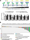
The GlcNAc-branching pathway in ocular surface epithelia. a, branches in complex-type N-glycans are processed through a series of enzymes, including MGAT1, MAN2A1, MAN2A2, MGAT2, MGAT4A, MGAT4B, and MGAT5 in the medial Golgi compartment. Gene expression of all of these enzymes was confirmed by qPCR in stratified human corneal and conjunctival cell lines, primary corneal epithelial cells, and native tissue (impression cytology) from human conjunctiva. b, the expression of N-glycan-processing genes in stratified cultures of human corneal epithelial cells was assessed by qPCR using a human glycosylation PCR array. The data show expression of B4GALT1 and B3GNT8, enzymes involved in the synthesis of poly-N-acetyllactosamine. In these experiments, negative or positive ΔΔCT values indicate, respectively, whether expression of the gene of interest is higher or lower than the reference gene MGAT1. Results in a represent three independent experiments performed in duplicate or triplicate. Data are represented as the mean ± S.D. Significance was determined using one-way analysis of variance with Tukey's post hoc test. The array in b was repeated twice with independently isolated RNA pooled from three tissue culture plates. *, p < 0.05; **, p < 0.01; ns, nonsignificant. GalT, galactosyltransferase.
Figure 3.
Figure 3.
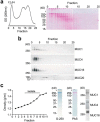
Purification of high-molecular-weight transmembrane mucins from stratified human corneal epithelial cells. An established two-step isolation procedure using size-exclusion chromatography and isopycnic density centrifugation was employed to purify transmembrane mucins. a, ∼10 mg of cell extract was applied to a Sepharose CL-4B column (1 × 30 cm) and eluted with PBS, pH 7.5. Fractions were evaluated for glycoprotein content using PAS staining. The asterisk indicates the stacking gel. b, by Western blotting, the high-molecular-weight fractions F1-F4 contained MUC1, MUC4, and MUC16 but not the smaller MUC20 transmembrane mucin. c, these fractions were pooled and subjected to isopycnic density centrifugation. Fractions at a buoyant density range of 1.26–1.43 g/ml tested positive for MUC1, MUC4, and MUC16 and were combined for N-glycan sequencing analyses. Coomassie G-250 staining revealed the presence of some low-molecular-weight bands in the mucin isolate; however, these bands were not glycosylated, as shown by PAS staining.
Figure 4.
Figure 4.
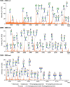
MALDI-TOF profile of N-glycans from transmembrane mucins isolated from human corneal epithelial cells. The N-linked glycans were released enzymatically by PNGase F, permethylated, and profiled by MALDI-TOF mass spectrometry. Magnified portions at m/z = 1500–2500, 2500–3500, and 3500–5500 are shown. Putative structures are assigned based on compositional information and known biosynthetic pathways. Most of the N-glycans on corneal mucins have compositions consistent with bi-, tri-, and tetra-antennary complex glycans, carrying N-acetyllactosamine residues and high mannose structures. All molecular ions detected are present in the form of [M + Na]+.
Figure 5.
Figure 5.
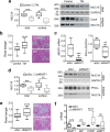
N-Glycosylation regulated MUC16 stability and the integrity of the corneal epithelial glycocalyx. a, tunicamycin (TM, 10 μg/ml) was added for the last 3 days of culture to inhibit synthesis of N-glycans in stratified human corneal epithelial cells. At the end of the incubation, cell-surface proteins were biotinylated at 4 °C, and an aliquot of ∼20% of the total cell lysate was stored before pulldown. The biotinylated proteins were isolated using NeutrAvidin™. Under these conditions, tunicamycin decreased MUC16 levels both in the cell extracts and on the plasma membrane. Inhibition of N-glycosylation by tunicamycin in these experiments was evaluated using ConA. b, Rose bengal penetrance into epithelial cells significantly increased after treatment with tunicamycin. c, cultures of human corneal epithelial cells were transfected with either non-targeting scramble control (siScr) or MGAT1-targeting siRNA (siMGAT1). Reduction in the expression of MGAT1 mRNA and N-glycan GlcNAc-branching was determined by qPCR and PHA-L (P. vulgaris leucoagglutinin) blot analysis, respectively. d, similar to tunicamycin, siRNA down-regulation of MGAT1 reduced the total and cell-surface levels of MUC16 protein. e, suppression of MGAT1 by siRNA was associated with an increase in Rose bengal penetrance into epithelial cells. f, the levels of MGAT1, BiP, CHOP, and sXBP1 mRNA were determined by qPCR. Results in a–f represent three independent experiments performed in triplicate. The box and whisker plots show the 25 and 75 percentiles (box) and the median and the minimum and maximum data values (whiskers). Significance was determined using Student's t test. **, p < 0.01; ***, p < 0.001; ****, p < 0.0001; ns, nonsignificant.
Figure 6.
Figure 6.
Galectin-3 interaction with MUC16 and its retention on the cell surface were N-glycan-dependent. a, 2-fold serial dilutions of rhGal-3 were applied individually to a nitrocellulose membrane in a slot-blot apparatus. Membranes were subsequently incubated with denatured cell lysates treated with or without PNGase F for 3 h at 37 °C. The enzymatic release of N-glycans significantly reduced the binding activity of MUC16 toward immobilized rhGal-3. Removal of N-glycans after PNGase F treatment was evaluated using ConA. b, treatment of stratified human corneal epithelial cells with PNGase F for 24 h significantly increased the amount of galectin-3 present in the cell culture medium. Relative amounts of N-glycans in biotinylated cell-surface proteins were evaluated using ConA. c, stratified human corneal epithelial cells were cultured in medium containing 1–10 μg/ml tunicamycin (TM) for the last 3 days of culture. Cells were then surface-labeled with biotin at 4 °C and pulled down using NeutrAvidin™. Galectin-3 was detected by Western blotting. d, cultures of human corneal epithelial cells were transfected with either non-targeting scramble control (siScr) or MGAT1-targeting siRNA (siMGAT1). As observed by cell-surface biotinylation and Western blotting, the knockdown of MGAT1 decreased the abundance of cell-surface galectin-3. Results in a and c represent at least three independent experiments. Results in b and d represent three independent experiments performed in triplicate. Data in c are represented as the mean ± S.D. The box and whisker plot show the 25 and 75 percentiles (box) and the median and the minimum and maximum data values (whiskers). Significance was determined using Student's t test (b and d) and one-way analysis of variance with Tukey's post hoc test (c). **, p < 0.01; ***, p < 0.001; ****, p < 0.0001; ns, nonsignificant.

Similar articles

Cited by

References

    1. McGuckin M. A., Lindén S. K., Sutton P., and Florin T. H. (2011) Mucin dynamics and enteric pathogens. Nat. Rev. Microbiol. 9, 265–278 - PubMed
    1. Corfield A. P. (2015) Mucins: a biologically relevant glycan barrier in mucosal protection. Biochim. Biophys. Acta 1850, 236–252 - PubMed
    1. Hilkens J., Ligtenberg M. J., Vos H. L., and Litvinov S. V. (1992) Cell membrane-associated mucins and their adhesion-modulating property. Trends Biochem. Sci. 17, 359–363 - PubMed
    1. Wesseling J., van der Valk S. W., Vos H. L., Sonnenberg A., and Hilkens J. (1995) Episialin (MUC1) overexpression inhibits integrin-mediated cell adhesion to extracellular matrix components. J. Cell Biol. 129, 255–265 - PMC - PubMed
    1. Bafna S., Kaur S., and Batra S. K. (2010) Membrane-bound mucins: the mechanistic basis for alterations in the growth and survival of cancer cells. Oncogene 29, 2893–2904 - PMC - PubMed