. 2017 Jun 23;292(25):10549-10563.
doi: 10.1074/jbc.M117.791707. Epub 2017 May 9.

Structure-function analysis of RBP-J-interacting and tubulin-associated (RITA) reveals regions critical for repression of Notch target genes

Affiliations

Structure-function analysis of RBP-J-interacting and tubulin-associated (RITA) reveals regions critical for repression of Notch target genes

Nassif Tabaja et al. J Biol Chem. .

Abstract

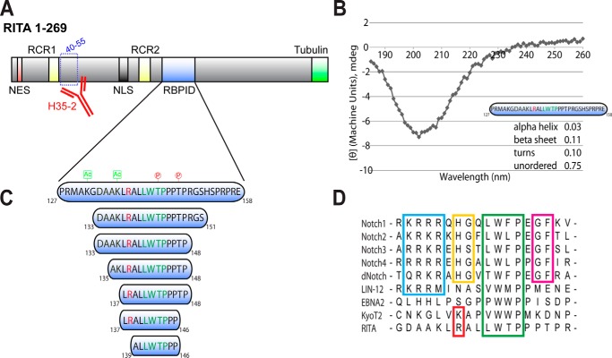
The Notch pathway is a cell-to-cell signaling mechanism that is essential for tissue development and maintenance, and aberrant Notch signaling has been implicated in various cancers, congenital defects, and cardiovascular diseases. Notch signaling activates the expression of target genes, which are regulated by the transcription factor CSL (CBF1/RBP-J, Su(H), Lag-1). CSL interacts with both transcriptional corepressor and coactivator proteins, functioning as both a repressor and activator, respectively. Although Notch activation complexes are relatively well understood at the structural level, less is known about how CSL interacts with corepressors. Recently, a new RBP-J (mammalian CSL ortholog)-interacting protein termed RITA has been identified and shown to export RBP-J out of the nucleus, thereby leading to the down-regulation of Notch target gene expression. However, the molecular details of RBP-J/RITA interactions are unclear. Here, using a combination of biochemical/cellular, structural, and biophysical techniques, we demonstrate that endogenous RBP-J and RITA proteins interact in cells, map the binding regions necessary for RBP-J·RITA complex formation, and determine the X-ray structure of the RBP-J·RITA complex bound to DNA. To validate the structure and glean more insights into function, we tested structure-based RBP-J and RITA mutants with biochemical/cellular assays and isothermal titration calorimetry. Whereas our structural and biophysical studies demonstrate that RITA binds RBP-J similarly to the RAM (RBP-J-associated molecule) domain of Notch, our biochemical and cellular assays suggest that RITA interacts with additional regions in RBP-J. Taken together, these results provide molecular insights into the mechanism of RITA-mediated regulation of Notch signaling, contributing to our understanding of how CSL functions as a transcriptional repressor of Notch target genes.

Keywords: Notch pathway; X-ray crystallography; gene regulation; isothermal titration calorimetry (ITC); protein-protein interaction; signal transduction; transcription corepressor; transcription factor.


Conflict of interest statement

The authors declare that they have no conflicts of interest with the contents of this article

Figures

Figure 1.
Figure 1.
Overview of Notch signaling. A, Notch signaling occurs between neighboring cells, in which interactions on the cell surface between a Notch receptor and a DSL ligand result in cleavage of Notch and release of its intracellular domain (NICD), which subsequently transits to the nucleus. B, in the absence of a Notch signal, CSL can bind corepressors, such as RITA, to repress transcription from Notch target genes. RITA binding to CSL can also cause CSL to be exported out of the nucleus. Upon activation of the Notch signaling pathway, NICD and MAM (Mastermind) form a ternary complex with CSL that activates transcription from Notch target genes. C, structure of the CSL·NICD·MAM ternary complex bound to DNA (PDB entry 2FO1). The structural core of CSL is composed of three domains (NTD, BTD, and CTD), which are colored cyan, green, and orange, respectively. A β-strand that makes hydrogen-bonding interactions with all three domains is colored magenta. The RAM and ANK domains of NICD are colored red and yellow, respectively. MAM and DNA are colored gray. D, domain schematics are colored similarly to the structure.
Figure 2.
Figure 2.
RITA domain schematic, secondary structure analysis, and sequence alignment with other CSL binding partners. A, RITA is a multidomain protein containing an N-terminal NES (red), RITA conserved repeats (RCR1/RCR2; yellow), NLS (gray), the RBPID (blue), and a C-terminal tubulin-interacting domain (green). As revealed by epitope mapping, the monoclonal antibody H35-2 (red) recognizes amino acids 40–55 of human RITA. B, far-UV CD spectra (wavelengths 185–200 nm) for the RBPID of RITA (amino acids 127–158). The RBPID consists of mostly random coil, as indicated by the minimum at 200 nm. Secondary structure was determined using Dichroweb and CDSSTR with reference set 7. The normalized root mean square deviation parameter value for the RITA CD data is 0.038. C, schematic representation of the RITA constructs used in this study with the ϕWϕP motif colored green and the arginine implicated in salt-bridge formation with Glu-260 of RBP-J colored red. Previously identified post-translational modifications within the RBPID are shown; lysine acetylation sites are colored green, and threonine phosphorylation sites are colored red. D, sequence alignment of coregulators that bind the BTD of CSL, including the RAM domains of human Notch1–4, the RAM domains of fly (dNotch) and worm (LIN-12) Notch receptors, the viral coactivator EBNA2, and the corepressor KyoT2. Boxed in blue, the RAM basic motif; boxed in yellow, the HG dipeptide motif; boxed in green, the ϕWϕP motif; boxed in magenta, the GF dipeptide motif; boxed in red, the basic residues of KyoT2 and RITA implicated in salt-bridge formation with RBP-J.
Figure 3.
Figure 3.
Endogenous RITA and RBP-J interact in cells. A, detection of endogenous RITA protein in HeLa cells (left) and HEK-293 cells (right). The indicated amounts of whole-cell lysates were used for Western blotting (WB). Membranes were incubated with the anti-RITA hybridoma supernatant H35-2. B, RITA expression levels in different human cell lines. Short exposure (top) and long exposure (middle) are shown. Expression of tubulin served as a loading control (bottom). Membranes were incubated with the anti-RITA hybridoma supernatant H35-2 (top and middle) or an anti-tubulin antibody (bottom). C, coimmunoprecipitations (IP) of endogenous RBP-J with RITA in HeLa cells. RBP-J was coimmunoprecipitated with RITA-specific hybridoma supernatants H35-1 (lane 1), H35-2 (lane 2), and H35-9 (lane 3) but not with an IgG control (lane 4). Purified RBP-J protein served as a positive control for Western blotting (lane 6). *, heavy chain of the antibodies.
Figure 4.
Figure 4.
Mapping the minimal RBP-J binding region of RITA within cells. A, schematic representation of RITA deletion constructs used for analysis of subcellular localization and interactions with endogenous RBP-J. Tubulin, tubulin-binding region. B, subcellular localization of GFP-RITA fusion proteins used for coimmunoprecipitation experiments. RITA(WT) and RITA(Δ128–156) show predominant tubulin association due to their tubulin binding region and rapid nucleocytoplasmic shuttling (19). RITA(156–296) is also located at tubulin fibers, confirming the tubulin binding region at the C terminus of RITA. RITA(83–173) and RITA(66–173) show predominantly nuclear localization, confirming the NLS within RITA. RITA(120–161) and RITA(106–173) show equal distribution within the cell. GFP localization served as a control. HeLa cells were transfected with the indicated GFP fusion constructs. 24 h after transfection, the living cells were imaged by fluorescence microscopy. Scale bar, 10 μm. C, coimmunoprecipitations (IP) of RBP-J with RITA deletion constructions. Top, RBP-J interacts with RITA(WT) (lane 1), RITA(83–173) (lane 3), RITA(106–173) (lane 4), and RITA(66–173) (lane 5) but not with RITA(156–269) (lane 2), RITA(Δ128–165) (lane 6), and RITA(120–161) (lane 7). Expression of RITA proteins (middle) and endogenous RBP-J (bottom) was verified by Western blotting (WB). Coimmunoprecipitations were performed 24 h after transfection of the indicated GFP-RITA fusions. *, heavy chain of anti-GFP antibody used for immunoprecipitation of RITA proteins.
Figure 5.
Figure 5.
Binding analysis of RBP-J/RITA interactions by ITC. Shown are representative thermograms from individual ITC experiments with various constructs of RBP-J and RITA. All ITC experiments were conducted with CSL in the cell at ∼20–25 μm and RITA in the syringe at ∼200–250 μm. Experimental temperature was set at 25 °C, and experiments were performed in triplicate (n = 3). A, a RITA construct (residues 106–173) that corresponds to the region necessary to interact with RBP-J binds with ∼1 μm affinity. B, a RITA construct (residues 127–158) that corresponds to the RBPID also binds with ∼1 μm affinity. C and D, the BTD and BTD-CTD constructs of RBP-J bind RITA(127–158) with ∼2 μm affinity. E, further truncation of the RBPID (residues 139–146) results in a significant loss of binding to RBP-J. F, ΔCp analysis of RBP-J/RITA interactions. ITC experiments were performed at 5, 15, 25, and 35 °C. The average change in Gibbs free energy (ΔG0), enthalpy (ΔH0), and entropy (−TΔS0) were plotted as a function of temperature.
Figure 6.
Figure 6.
High-resolution structure of the RBP-J·RITA corepressor complex bound to DNA. A, the X-ray structure of RBP-J·RITA·DNA (PDB entry 5EG6) was determined to 2.1 Å resolution. Shown are a ribbon and surface representation of the complex structure with the NTD, BTD, and CTD colored cyan, green, and orange, respectively; RITA is colored yellow. B, structural overlay of CTD domains from the RBP-J·RITA·DNA structure determined here with the CTD from the previously published RBP-J/DNA structure (PDB entry 3IAG), highlighting the 10.5-Å rigid body shift of the CTD of RBP-J when bound to RITA. C, enlarged view of RITA binding the BTD of RBP-J, emphasizing the ϕWϕP motif. RITA is shown in a stick representation with corresponding 2FoFc electron density contoured at 1σ. D, salt bridge formed between Arg-138 of RITA (yellow) and Glu-259 and Glu-260 of RBP-J (green). E, structural alignment of BTD-binding proteins, including RITA in yellow (PDB entry 5EG6), KyoT2 in blue (PDB entry 42JX), worm RAM in pink (PDB entry 3BRD), and human RAM in red (PDB entry 3V79). Cα traces are shown with the side chains of the ϕWϕP motifs depicted as sticks. F, location of the mutation sites (Phe-261, Val-263, Ala-284, and Gln-333) within the BTD that affect RAM binding and were used in this study.
Figure 7.
Figure 7.
Cellular reporter assays of RITA-mediated repression in the context of CSL mutants. RBP-J null MEFs were transduced with a retrovirus encoding either wild-type or mutant RBP-J constructs. To activate and readout Notch signaling, cells were transfected with a construct that expresses an activated form of the Notch1 receptor (NICD1) and the 4×CBS reporter, which has four CSL-binding sites upstream of the firefly luciferase gene. To assay for RITA-mediated repression, cells were cotransfected with increasing amounts of a construct that expresses RITA: 0 ng (−), 50 ng (+), 100 ng (++), 200 ng (+++), or 400 ng (++++). Experiments were performed in triplicate, and the error bars represent S.E. A, RITA represses Notch reporter activity in a dose-dependent manner. -Fold activation is relative to luciferase activity from control cells not transfected with NICD1. B, plot shows reduced reporter activity for RBP-J mutants (F261A, V263A, A284V, and Q333A). -Fold activation is relative to luciferase activity from control cells not transfected with NICD1. C–F, plots show RITA-mediated repression for the RBP-J mutants compared with wild type. Data are normalized to cells with NICD1, but without RITA, and shown as relative activity. Statistical significance was determined by unpaired t test. *, p ≤ 0.05; **, p ≤ 0.01; ns, not significant.
Figure 8.
Figure 8.
Cellular reporter assays of RITA-mediated repression in the context of RITA mutants. Cellular reporter assays in retrovirally transduced MEFs were performed similarly as described for Fig. 5. A, plot shows relative repression activity of RITA (residues 106–173), which contains the RBPID, compared with full-length RITA. B, plot shows reduced, but not completely abolished, RITA-mediated repression of the reporter for the RITA mutant WTP/AAA, which mutates the ϕWϕP motif. C, plot shows reduced reporter activity, compared with wild type, for the construct RITAΔNES, which deletes the nuclear export sequence of RITA. D, plot shows reduced reporter activity, compared with wild type, for the construct RITAΔNLS, containing a non-functional NLS. Error bars, S.E. Statistical significance was determined by unpaired t test. *, p ≤ 0.05; **, p ≤ 0.01; ns, not significant.

Similar articles

Cited by

References

    1. Bray S. J. (2016) Notch signalling in context. Nat. Rev. Mol. Cell Biol. 17, 722–735 - PubMed
    1. Swiatek P. J., Lindsell C. E., del Amo F. F., Weinmaster G., and Gridley T. (1994) Notch1 is essential for postimplantation development in mice. Genes Dev. 8, 707–719 - PubMed
    1. Gridley T. (2003) Notch signaling and inherited disease syndromes. Hum. Mol. Genet. 12, R9–R13 - PubMed
    1. Koch U., and Radtke F. (2010) Notch signaling in solid tumors. Curr. Top. Dev. Biol. 92, 411–455 - PubMed
    1. Ntziachristos P., Lim J. S., Sage J., and Aifantis I. (2014) From fly wings to targeted cancer therapies: a centennial for notch signaling. Cancer Cell 25, 318–334 - PMC - PubMed

Publication types

MeSH terms