

Structural Basis for Linezolid Binding Site Rearrangement in the Staphylococcus aureus Ribosome
- PMID: 28487427
- PMCID: PMC5424203
- DOI: 10.1128/mBio.00395-17
Structural Basis for Linezolid Binding Site Rearrangement in the Staphylococcus aureus Ribosome
Abstract
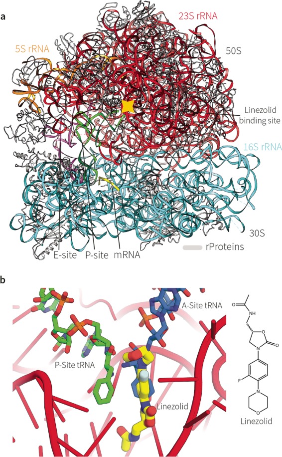
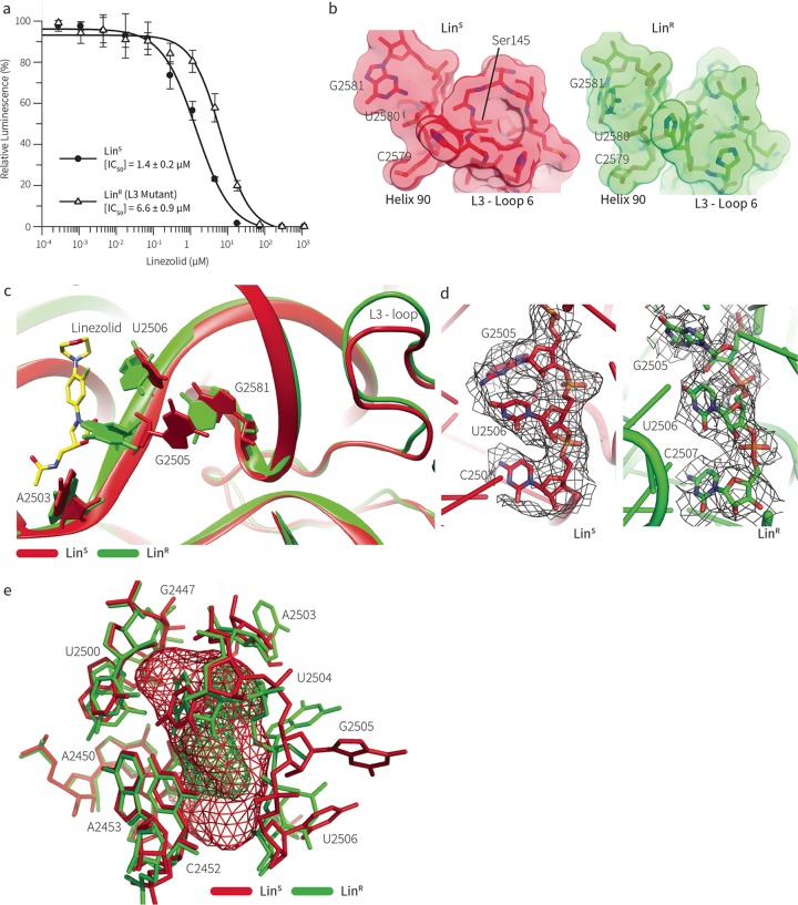
An unorthodox, surprising mechanism of resistance to the antibiotic linezolid was revealed by cryo-electron microscopy (cryo-EM) in the 70S ribosomes from a clinical isolate of Staphylococcus aureus This high-resolution structural information demonstrated that a single amino acid deletion in ribosomal protein uL3 confers linezolid resistance despite being located 24 Å away from the linezolid binding pocket in the peptidyl-transferase center. The mutation induces a cascade of allosteric structural rearrangements of the rRNA that ultimately results in the alteration of the antibiotic binding site.IMPORTANCE The growing burden on human health caused by various antibiotic resistance mutations now includes prevalent Staphylococcus aureus resistance to last-line antimicrobial drugs such as linezolid and daptomycin. Structure-informed drug modification represents a frontier with respect to designing advanced clinical therapies, but success in this strategy requires rapid, facile means to shed light on the structural basis for drug resistance (D. Brown, Nat Rev Drug Discov 14:821-832, 2015, https://doi.org/10.1038/nrd4675). Here, detailed structural information demonstrates that a common mechanism is at play in linezolid resistance and provides a step toward the redesign of oxazolidinone antibiotics, a strategy that could thwart known mechanisms of linezolid resistance.
Keywords: antibiotic resistance; ribosomal mutations; staphylococcus.
Copyright © 2017 Belousoff et al.
Figures
Similar articles
-
Resistance to linezolid in Staphylococcus aureus by mutation, modification, and acquisition of genes.J Antibiot (Tokyo). 2025 Jan;78(1):4-13. doi: 10.1038/s41429-024-00778-4. Epub 2024 Oct 17. J Antibiot (Tokyo). 2025. PMID: 39420155 Free PMC article. Review.
-
A Structurally Characterized Staphylococcus aureus Evolutionary Escape Route from Treatment with the Antibiotic Linezolid.Microbiol Spectr. 2022 Aug 31;10(4):e0058322. doi: 10.1128/spectrum.00583-22. Epub 2022 Jun 23. Microbiol Spectr. 2022. PMID: 35736238 Free PMC article.
-
Exit tunnel modulation as resistance mechanism of S. aureus erythromycin resistant mutant.Sci Rep. 2019 Aug 7;9(1):11460. doi: 10.1038/s41598-019-48019-1. Sci Rep. 2019. PMID: 31391518 Free PMC article.
-
Linezolid-dependent function and structure adaptation of ribosomes in a Staphylococcus epidermidis strain exhibiting linezolid dependence.Antimicrob Agents Chemother. 2014 Aug;58(8):4651-6. doi: 10.1128/AAC.02835-14. Epub 2014 Jun 2. Antimicrob Agents Chemother. 2014. PMID: 24890589 Free PMC article.
-
Resistance to linezolid caused by modifications at its binding site on the ribosome.Antimicrob Agents Chemother. 2012 Feb;56(2):603-12. doi: 10.1128/AAC.05702-11. Epub 2011 Dec 5. Antimicrob Agents Chemother. 2012. PMID: 22143525 Free PMC article. Review.
Cited by
-
Context-Specific Action of Ribosomal Antibiotics.Annu Rev Microbiol. 2018 Sep 8;72:185-207. doi: 10.1146/annurev-micro-090817-062329. Epub 2018 Jun 15. Annu Rev Microbiol. 2018. PMID: 29906204 Free PMC article. Review.
-
Structural Studies Reveal the Role of Helix 68 in the Elongation Step of Protein Biosynthesis.mBio. 2022 Apr 26;13(2):e0030622. doi: 10.1128/mbio.00306-22. Epub 2022 Mar 29. mBio. 2022. PMID: 35348349 Free PMC article.
-
Resistance to linezolid in Staphylococcus aureus by mutation, modification, and acquisition of genes.J Antibiot (Tokyo). 2025 Jan;78(1):4-13. doi: 10.1038/s41429-024-00778-4. Epub 2024 Oct 17. J Antibiot (Tokyo). 2025. PMID: 39420155 Free PMC article. Review.
-
Strategies to Improve the Potency of Oxazolidinones towards Bacterial Biofilms.Chem Asian J. 2022 Jun 1;17(11):e202200201. doi: 10.1002/asia.202200201. Epub 2022 Apr 13. Chem Asian J. 2022. PMID: 35352479 Free PMC article. Review.
-
Linezolid: a review of its properties, function, and use in critical care.Drug Des Devel Ther. 2018 Jun 18;12:1759-1767. doi: 10.2147/DDDT.S164515. eCollection 2018. Drug Des Devel Ther. 2018. PMID: 29950810 Free PMC article. Review.
References
-
- Bashan A, Agmon I, Zarivach R, Schluenzen F, Harms J, Berisio R, Bartels H, Franceschi F, Auerbach T, Hansen HA, Kossoy E, Kessler M, Yonath A. 2003. Structural basis of the ribosomal machinery for peptide bond formation, translocation, and nascent chain progression. Mol Cell 11:91–102. doi:10.1016/S1097-2765(03)00009-1. - DOI - PubMed
MeSH terms
Substances
Associated data
- Actions
LinkOut - more resources
Full Text Sources
Other Literature Sources
Medical
Molecular Biology Databases
