

Auditory Target and Novelty Processing in Patients with Unilateral Hippocampal Sclerosis: A Current-Source Density Study
- PMID: 28487515
- PMCID: PMC5431625
- DOI: 10.1038/s41598-017-01531-8
Auditory Target and Novelty Processing in Patients with Unilateral Hippocampal Sclerosis: A Current-Source Density Study
Abstract
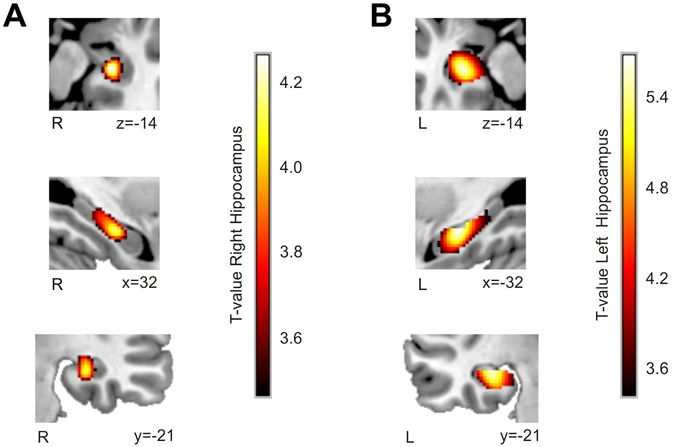
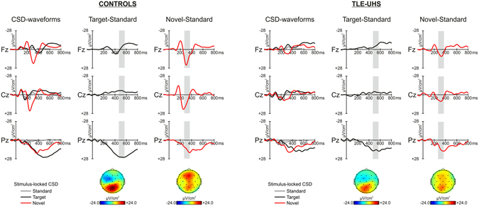
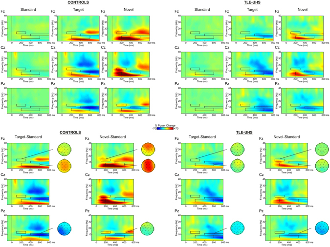
The capacity to respond to novel events is crucial for adapting to the constantly changing environment. Here, we recorded 29-channel Event Related Brain Potentials (ERPs) during an active auditory novelty oddball paradigm and used for the first time Current Source Density-transformed Event Related Brain Potentials and associated time-frequency spectra to study target and novelty processing in a group of epileptic patients with unilateral damage of the hippocampus (N = 18) and in healthy matched control participants (N = 18). Importantly, we used Voxel-Based Morphometry to ensure that our group of patients had a focal unilateral damage restricted to the hippocampus and especially its medial part. We found a clear deficit for target processing at the behavioral level. In addition, compared to controls, our group of patients presented (i) a reduction of theta event-related synchronization (ERS) for targets and (ii) a reduction and delayed P3a source accompanied by reduced theta and low-beta ERS and alpha event-related synchronization (ERD) for novel stimuli. These results suggest that the integrity of the hippocampus might be crucial for the functioning of the complex cortico-subcortical network involved in the detection of novel and target stimuli.
Conflict of interest statement
The authors declare no competing interests.
Figures
Similar articles
-
Role of acetylcholine and serotonin in novelty processing using an oddball paradigm.Behav Brain Res. 2017 Jul 28;331:199-204. doi: 10.1016/j.bbr.2017.05.031. Epub 2017 May 13. Behav Brain Res. 2017. PMID: 28511977
-
Neuromagnetic evidence for hippocampal modulation of auditory processing.Neuroimage. 2016 Jan 1;124(Pt A):256-266. doi: 10.1016/j.neuroimage.2015.09.006. Epub 2015 Sep 10. Neuroimage. 2016. PMID: 26363346
-
Regional and inter-regional theta oscillation during episodic novelty processing.Brain Cogn. 2014 Oct;90:70-5. doi: 10.1016/j.bandc.2014.06.009. Epub 2014 Jul 9. Brain Cogn. 2014. PMID: 25014407
-
[Progress of study in auditory event-related potentials of novel events].Sheng Wu Yi Xue Gong Cheng Xue Za Zhi. 2007 Jun;24(3):705-8. Sheng Wu Yi Xue Gong Cheng Xue Za Zhi. 2007. PMID: 17713294 Review. Chinese.
-
Anatomic bases of event-related potentials and their relationship to novelty detection in humans.J Clin Neurophysiol. 1998 Jan;15(1):3-13. doi: 10.1097/00004691-199801000-00003. J Clin Neurophysiol. 1998. PMID: 9502509 Review. No abstract available.
Cited by
-
Event-related modulation of alpha rhythm explains the auditory P300-evoked response in EEG.Elife. 2023 Dec 1;12:RP88367. doi: 10.7554/eLife.88367. Elife. 2023. PMID: 38038725 Free PMC article.
-
Effects of aging on neural processing during an active listening task.PLoS One. 2022 Sep 7;17(9):e0273304. doi: 10.1371/journal.pone.0273304. eCollection 2022. PLoS One. 2022. PMID: 36070253 Free PMC article.
References
Publication types
MeSH terms
LinkOut - more resources
Full Text Sources
Other Literature Sources
