. 2017 May 9;7(1):1597.
doi: 10.1038/s41598-017-01716-1.

Retinal Degeneration In A Mouse Model Of CLN5 Disease Is Associated With Compromised Autophagy

Affiliations

Retinal Degeneration In A Mouse Model Of CLN5 Disease Is Associated With Compromised Autophagy

Henri Leinonen et al. Sci Rep. .

Abstract

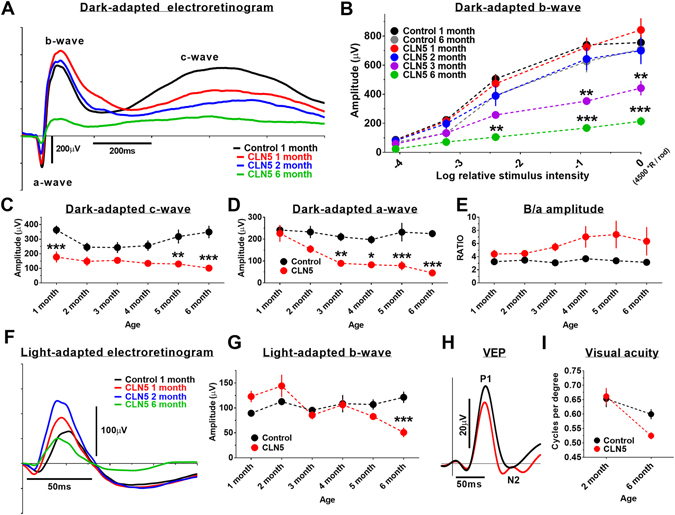
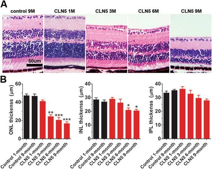
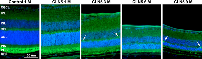
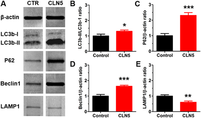
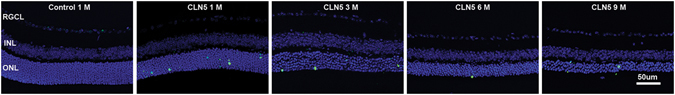
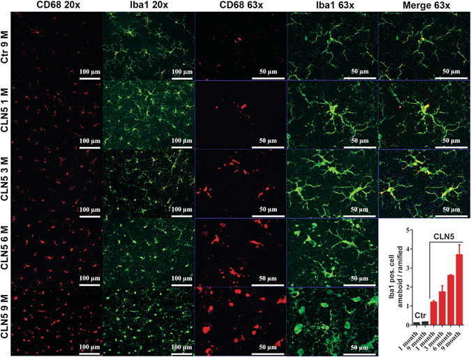
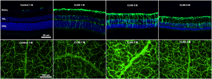
The Finnish variant of late infantile neuronal ceroid lipofuscinosis (CLN5 disease) belongs to a family of neuronal ceroid lipofuscinosis (NCLs) diseases. Vision loss is among the first clinical signs in childhood forms of NCLs. Mutations in CLN5 underlie CLN5 disease. The aim of this study was to characterize how the lack of normal functionality of the CLN5 protein affects the mouse retina. Scotopic electroretinography (ERG) showed a diminished c-wave amplitude in the CLN5 deficient mice already at 1 month of age, indicative of pathological events in the retinal pigmented epithelium. A- and b-waves showed progressive impairment later from 2 and 3 months of age onwards, respectively. Structural and immunohistochemical (IHC) analyses showed preferential damage of photoreceptors, accumulation of autofluorescent storage material, apoptosis of photoreceptors, and strong inflammation in the CLN5 deficient mice retinas. Increased levels of autophagy-associated proteins Beclin-1 and P62, and increased LC3b-II/LC3b-I ratio, were detected by Western blotting from whole retinal extracts. Photopic ERG, visual evoked potentials, IHC and cell counting indicated relatively long surviving cone photoreceptors compared to rods. In conclusion, CLN5 deficient mice develop early vision loss that reflects the condition reported in clinical childhood forms of NCLs. The vision loss in CLN5 deficient mice is primarily caused by photoreceptor degeneration.


Conflict of interest statement

The authors declare that they have no competing interests.

Figures

Figure 1
Figure 1
Progressive decline in retinal function and cortical visual acuity in CLN5 deficient mice. (A) group averaged band- filtered (70–150 Hz) scotopic ERG waveforms (at log 0) in 1-month-old control and CLN5 deficient mice (groups sizes: control 1 month, n = 12; control 2 month, n = 10, control 3–6 month, n = 6; CLN5 1 month, n = 9; CLN5 2–6 month, n = 6–8). (B) scotopic b-wave amplitudes over age. Note that all age points are not shown for clarity of presentation. The maximum stimulus intensity (log 0) yielded ~4500 photoisomerizations per rod. (C,D) scotopic c- and a-wave amplitudes (at log 0) in control and CLN5 deficient mice over age, respectively. (E) scotopic b-wave/a-wave amplitude ratio at log 0 flash. (F) group averaged band- filtered (70–150 Hz) photopic ERG waveforms (at log 0) in 1-month-old control and 1-month, 2-month and 6-month-old CLN5 deficient mice. (G) photopic b-wave amplitudes (at log 0) in control and CLN5 deficient mice over age. (H) Representative group averaged VEP waveforms in response to middle size (0.192 CPD) pattern stimulus in 6-month-old mice. (I) Cortical visual acuity as determined by pattern VEPs (n = 11 per genotype). Bonferroni multiple comparison test: *p < 0.05, **p < 0.01, ***p < 0.001.
Figure 2
Figure 2
Robust photoreceptor death in CLN5 deficient mice. (A) Representative H&E stained retinal sections. At 1 month of age CLN5 deficient retina represents completely normal morphology. Also at 3 month of age the CLN5 deficient retina has normal appearance but the ONL is drastically thinned. The pathology progressed so that the POS started to become disorganized and disappeared, and at the late state of disease at 9 months of age even PIS died out. The inner retinal compartments remained normal appearance over the course of the follow up. (B) Statistical analysis of ONL, INL and IPL thicknesses (control 1 month, n = 4–5; control 9 month, n = 4; CLN5 1 month, n = 4; CLN5 3 month, n = 5; CLN5 6 month, n = 6–7; CLN5 9 month, n = 6) showed that ONL was primarily affected in CLN5 deficient mice retinas with early onset whereas INL and IPL showed slowly progressive thinning. Tukey’s posthoc test: *p < 0.05, **p < 0.01, ***p < 0.001. RGC, retinal ganglion cells; IPL, inner plexiform layer; INL, inner nuclear layer; OPL, outer plexiform layer; ONL, outer nuclear layer; PIS, photoreceptor inner segments; POS, photoreceptor outer segments.
Figure 3
Figure 3
Increased autofluorescence (AF) in CLN5 deficient mouse retinas. Images were captured with a fluorescent microscope using auto-exposure. CLN5 deficient retinas seem to display stronger diffuse AF throughout the retina as compared to wild-type control. AF aggregates started to appear from 3 month of age onwards particularly into the OPL (arrows), which is more obvious in Supplementary Fig. 2. RGCL, retinal ganglion cell layer; IPL, inner plexiform layer; INL, inner nuclear layer; OPL, outer plexiform layer; ONL, outer nuclear layer; PIS, photoreceptor inner segments; POS, photoreceptor outer segments; RPE, retinal pigmented epithelium.
Figure 4
Figure 4
Altered autophagy in 6-month-old CLN5 deficient mouse retinas. (A) Representative immunoblots. (B) LC3-I to LC-II conversion was increased 1.3-fold in CLN5 deficient retinas (n = 10 per genotype). (C,D) P62 level was increased 2.3-fold, and Beclin-1 level 1.6-fold in CLN5 deficient retinas (n = 7 and 8 per genotype, respectively). (E) LAMP1 level was decreased in CLN5 deficient retinas and was 0.6-fold lower when compared to control levels (n = 8 per genotype). The protein level ratios in CLN5 deficient samples were compared to normalized control values in all analyses (B–E). *p < 0.05, **p < 0.01, p < 0.001; t-test.
Figure 5
Figure 5
Apoptotic photoreceptor nuclei in CLN5 deficient mice retinas. Representative fluorescent z-stack images of TUNEL staining (green) with DAPI counter stain (blue, cell nuclei). At 1 month of age apoptotic cells were frequent in CLN5 deficient mice samples but the amount decreased with diminishing amount of surviving photoreceptors (see Supplementary Fig. 3). Apoptotic cells were restricted to the ONL. RGCL, retinal gangion cell layer; INL, inner nuclear layer; ONL, outer nuclear layer.
Figure 6
Figure 6
Early microglial infiltration and activation in CLN5 deficient mice retinas. Layout of representative z-stack images taken at the vicinity of ONL are shown for control and different aged CLN5 deficient retinal whole mount samples (rows, horizontal). CD68 (red, macrophage marker) and Iba1 positive (green, microglia marker) cells are shown with two different magnifications vertically in columns. Already at 1 month of age the CLN5 deficient retinas showed remarkably increased CD68 and Iba1 positivity and microglia cell morphology was changing from ramified form (inactive microglia) to a more amoeboid (active microglia) form. This pathological process progressed so that at 9 month of age very few Iba1 positive cells had ramified appearance. CD68 positivity was localized to the microglial cell body. Down-right corner: amoeboid – ramified cell morphology ratio was estimated by manual counting from Iba1 positive cells at 8 retinal locations and averaged for illustration.
Figure 7
Figure 7
Müller cell gliosis and astrogliosis in CLN5 deficient mice retinas. Upper row: representative fluorescent z-stack images are shown from anti-GFAP (green, macroglia) staining with a DAPI counterstain (blue, cell nuclei). Astrocytes localize at the retinal nerve fiber layer, above the RGCL, horizontally and are expressing GFAP little in control samples. Müller cells are vertically oriented in the retina and do not express GFAP once inactivated. Already at 1 month of age, astrocytes and Müller cells were strongly activated in CLN5 deficient mice as shown by intense GFAP positivity. GFAP positive staining increased until 3 month of age. Lower row: anti-GFAP staining of retinal whole mounts. RGCL, retinal ganglion cell layer; INL, inner nuclear layer; ONL, outer nuclear layer; GFAP, glial fibrillary acidid protein.

Similar articles

Cited by

References

    1. Haltia M. The neuronal ceroid-lipofuscinoses: from past to present. Biochim Biophys Acta. 2006;1762:850–6. doi: 10.1016/j.bbadis.2006.06.010. - DOI - PubMed
    1. Carcel-Trullols J, Kovacs AD, Pearce DA. Cell biology of the NCL proteins: What they do and don’t do. Biochim Biophys Acta. 2015;1852:2242–55. doi: 10.1016/j.bbadis.2015.04.027. - DOI - PubMed
    1. Kollmann K, et al. Cell biology and function of neuronal ceroid lipofuscinosis-related proteins - Biochim Biophys Acta. 2013;1832:1866–81. doi: 10.1016/j.bbadis.2013.01.019. - DOI - PubMed
    1. Collins J, et al. Batten disease: features to facilitate early diagnosis. Br J Ophthalmol. 2006;90:1119–24. doi: 10.1136/bjo.2006.091637. - DOI - PMC - PubMed
    1. Radke J, Stenzel W, Goebel HH. Human NCL Neuropathology. Biochim Biophys Acta. 2015;1852:2262–6. doi: 10.1016/j.bbadis.2015.05.007. - DOI - PubMed

Publication types

MeSH terms

Substances