

Effect of Drop Foot on Spatiotemporal, Kinematic, and Kinetic Parameters during Gait
- PMID: 28487621
- PMCID: PMC5405572
- DOI: 10.1155/2017/3595461
Effect of Drop Foot on Spatiotemporal, Kinematic, and Kinetic Parameters during Gait
Abstract
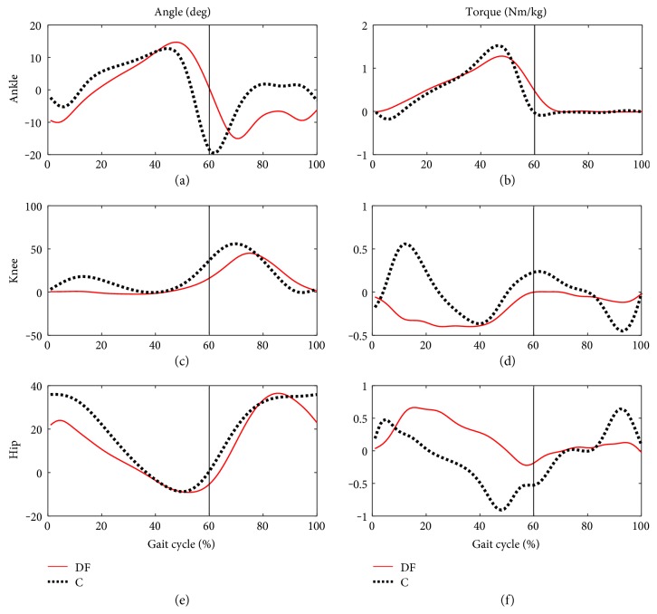
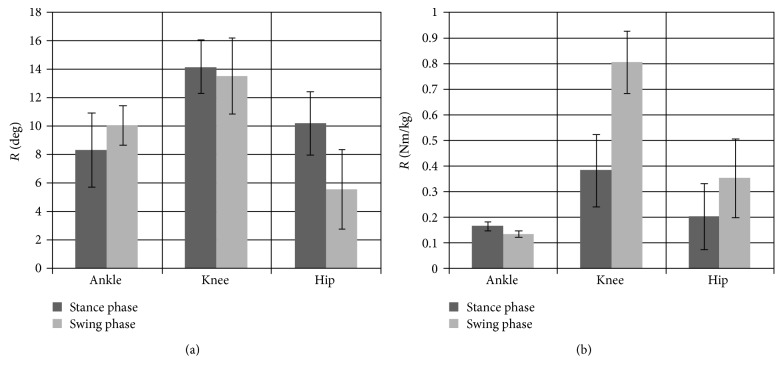
Background. The complexity of the structure and function of a living body can be affected by disorders and can cause various dysfunctions. Objective. The aim of this study was to determine compensatory mechanisms in subjects with drop foot during gait. Methods. The study evaluated 10 subjects with drop foot (DF) whose results were compared to a group of 10 healthy controls (C). Spatiotemporal, kinematic, and kinetic parameters during the gait cycle were collected using Vicon system synchronized with Kistler platforms. Results. Spatiotemporal, kinematic, and kinetic parameters were significantly different between the analysed groups. In the DF group, the subjects walked almost 47% slower and performed 60% less steps per minute compared to the C group. The main problem in the DF group was insufficient ankle dorsiflexion in the 0-10% of the gait cycle. Mean values in the groups during the first 10% of the gait cycle were as follows: DF (-10.42 ± 5.7°) and C (-2.37 ± 1.47°), which affected the substantial differences in the values of muscle torque: DF (0.2 ± 0.1 Nm/kg) and C (-0.26 ± 0.06 Nm/kg). Conclusions. Comparative analysis for joint angles and torques demonstrated that the mechanism of compensation is the most noticeable in the knee joint and less in the hip joint.
Figures
References
-
- Perry J. S. Incorporated. Normal and Pathological Function. Thorofare, NJ, USA: SLACK Incorporated; 1992. Gait analysis. - DOI
LinkOut - more resources
Full Text Sources
Other Literature Sources
Miscellaneous
