The G Protein-Coupled Receptor UT of the Neuropeptide Urotensin II Displays Structural and Functional Chemokine Features
- PMID: 28487672
- PMCID: PMC5403833
- DOI: 10.3389/fendo.2017.00076
The G Protein-Coupled Receptor UT of the Neuropeptide Urotensin II Displays Structural and Functional Chemokine Features
Abstract
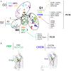
The urotensinergic system was previously considered as being linked to numerous physiopathological states, including atherosclerosis, heart failure, hypertension, pre-eclampsia, diabetes, renal disease, as well as brain vascular lesions. Thus, it turns out that the actions of the urotensin II (UII)/G protein-coupled receptor UT system in animal models are currently not predictive enough in regard to their effects in human clinical trials and that UII analogs, established to target UT, were not as beneficial as expected in pathological situations. Thus, many questions remain regarding the overall signaling profiles of UT leading to complex involvement in cardiovascular and inflammatory responses as well as cancer. We address the potential UT chemotactic structural and functional definition under an evolutionary angle, by the existence of a common conserved structural feature among chemokine receptorsopioïdergic receptors and UT, i.e., a specific proline position in the transmembrane domain-2 TM2 (P2.58) likely responsible for a kink helical structure that would play a key role in chemokine functions. Even if the last decade was devoted to the elucidation of the cardiovascular control by the urotensinergic system, we also attempt here to discuss the role of UII on inflammation and migration, likely providing a peptide chemokine status for UII. Indeed, our recent work established that activation of UT by a gradient concentration of UII recruits Gαi/o and Gα13 couplings in a spatiotemporal way, controlling key signaling events leading to chemotaxis. We think that this new vision of the urotensinergic system should help considering UT as a chemotactic therapeutic target in pathological situations involving cell chemoattraction.
Keywords: G protein-coupled receptor; UT; chemokine; migration; proline; urotensin II.
Figures


Similar articles
-
Signaling switch of the urotensin II vasosactive peptide GPCR: prototypic chemotaxic mechanism in glioma.Oncogene. 2015 Sep 24;34(39):5080-94. doi: 10.1038/onc.2014.433. Epub 2015 Jan 19. Oncogene. 2015. PMID: 25597409
-
Update on the urotensinergic system: new trends in receptor localization, activation, and drug design.Front Endocrinol (Lausanne). 2013 Jan 2;3:174. doi: 10.3389/fendo.2012.00174. eCollection 2012. Front Endocrinol (Lausanne). 2013. PMID: 23293631 Free PMC article.
-
Increased expression of urotensin II, urotensin II-related peptide and urotensin II receptor mRNAs in the cardiovascular organs of hypertensive rats: comparison with endothelin-1.Peptides. 2009 Jun;30(6):1124-9. doi: 10.1016/j.peptides.2009.02.009. Epub 2009 Feb 21. Peptides. 2009. PMID: 19463745
-
Emerging roles of urotensin-II in cardiovascular disease.Pharmacol Ther. 2004 Sep;103(3):223-43. doi: 10.1016/j.pharmthera.2004.07.004. Pharmacol Ther. 2004. PMID: 15464591 Review.
-
Role of urotensin II in health and disease.Am J Physiol Regul Integr Comp Physiol. 2010 May;298(5):R1156-72. doi: 10.1152/ajpregu.00706.2009. Am J Physiol Regul Integr Comp Physiol. 2010. PMID: 20421634 Review.
Cited by
-
Targeting the Urotensin II/UT G Protein-Coupled Receptor to Counteract Angiogenesis and Mesenchymal Hypoxia/Necrosis in Glioblastoma.Front Cell Dev Biol. 2021 Apr 14;9:652544. doi: 10.3389/fcell.2021.652544. eCollection 2021. Front Cell Dev Biol. 2021. PMID: 33937253 Free PMC article.
-
Serum Urotensin II Levels Are Elevated in Patients with Obstructive Sleep Apnea.Biomolecules. 2023 May 31;13(6):914. doi: 10.3390/biom13060914. Biomolecules. 2023. PMID: 37371494 Free PMC article.
-
Activation of the urotensin-II receptor by remdesivir induces cardiomyocyte dysfunction.Commun Biol. 2023 May 12;6(1):511. doi: 10.1038/s42003-023-04888-x. Commun Biol. 2023. PMID: 37173432 Free PMC article.
-
The network map of urotensin-II mediated signaling pathway in physiological and pathological conditions.J Cell Commun Signal. 2022 Dec;16(4):601-608. doi: 10.1007/s12079-022-00672-4. Epub 2022 Feb 16. J Cell Commun Signal. 2022. PMID: 35174439 Free PMC article.
-
Isoproterenol mechanisms in inducing myocardial fibrosis and its application as an experimental model for the evaluation of therapeutic potential of phytochemicals and pharmaceuticals.Animal Model Exp Med. 2025 Jan;8(1):67-91. doi: 10.1002/ame2.12496. Epub 2024 Dec 17. Animal Model Exp Med. 2025. PMID: 39690876 Free PMC article. Review.
References
-
- Bern HA, Lederis K. A reference preparation for the study of active substances in the caudal neurosecretory system of teleosts. J Endocrinol (1969) 45:Sul:xi–xii. - PubMed
-
- Coulouarn Y, Lihrmann I, Jegou S, Anouar Y, Tostivint H, Beauvillain JC, et al. Cloning of the cDNA encoding the urotensin II precursor in frog and human reveals intense expression of the urotensin II gene in motoneurons of the spinal cord. Proc Natl Acad Sci U S A (1998) 95:15803–8.10.1073/pnas.95.26.15803 - DOI - PMC - PubMed
Publication types
LinkOut - more resources
Full Text Sources
Other Literature Sources

