Case Reports
. 2017:2017:8562050.
doi: 10.1155/2017/8562050. Epub 2017 Apr 9.

Biofilm Analysis of Retrieved Dental Implants after Different Peri-Implantitis Treatments

Affiliations
Case Reports

Biofilm Analysis of Retrieved Dental Implants after Different Peri-Implantitis Treatments

Thaise C Geremias et al. Case Rep Dent. 2017.

Abstract

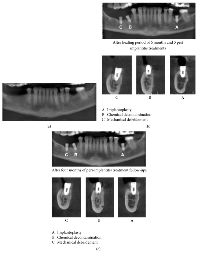
The aim of the current study was to analyse the planktonic growth of Streptococcus mutans on the surfaces of three implants retrieved after three different peri-implantitis treatments. Three implants from a male patient with high levels of bone loss were treated by mechanical debridement, chemical decontamination, and implantoplasty. After 4 months of follow-up, the implants were removed. The growth and biofilm formation were measured by spectrophotometry (OD630 nm) and scanning electron microscopy (SEM), after 48 hours of incubation. Results showed an average of Streptococcus mutans planktonic growth over the implants of 0.21 nm (mechanical debridement), 0.16 nm (chemical decontamination), and 0.15 nm (implantoplasty). Data were analysed by ANOVA and Tukey's test (p < 0.05 for chemical decontamination and implantoplasty). Implantoplasty and chemical decontamination showed the lowest levels of planktonic growth, indicating a possible influence of the modification procedures on the titanium surface on the initial biofilm attachment.


Figures

Figure 1
Figure 1
(a) Initial tomography, (b) 6 months' implant after placement and peri-implantitis treatments, (c) 4 months' after different peri-implantitis treatments. (A: implantoplasty, B: acid treatment, and C: debridement).
Figure 2
Figure 2
Peri-implantitis treatment. A: implantoplasty, B: acid treatment, and C: debridement.
Figure 3
Figure 3
Scanning electron microscopy (SEM) images of the titanium surface displaying topography features before and after the S. mutans biofilm formation ((A) implantoplasty, (B) acid treatment, and (C) debridement) (Magnification D8.6 ×1.0k 100 μm).
Figure 4
Figure 4
Biofilm formation on implantoplasty treatment surface (a). S. mutans planktonic growth in implants for 48 h in TSB (37°C), measured by spectrophotometer with an optical density of 630 nm (b) and table exhibiting mean values, according to the specific treatment (c).

References

    1. Albrektsson T., Buser D., Chen S. T., et al. Statements from the Estepona consensus meeting on peri-implantitis, February 2–4, 2012. Clinical Implant Dentistry and Related Research. 2012;14(6):781–782. doi: 10.1111/cid.12017. - DOI - PubMed
    1. Klinge B., Flemming T., Cosyn J., et al. The patient undergoing implant therapy. Summary and consensus statements. The 4th EAO Consensus Conference 2015. Clinical Oral Implants Research. 2015;26, supplement 1:64–67. doi: 10.1111/clr.12675. - DOI - PubMed
    1. Marsh P. D. Dental plaque: biological significance of a biofilm and community life-style. Journal of Clinical Periodontology. 2005;32(6):7–15. doi: 10.1111/j.1600-051x.2005.00790.x. - DOI - PubMed
    1. Teughels W., Van Assche N., Sliepen I., Quirynen M. Effect of material characteristics and/or surface topography on biofilm development. Clinical Oral Implants Research. 2006;17(supplement 2):68–81. doi: 10.1111/j.1600-0501.2006.01353.x. - DOI - PubMed
    1. Costerton J. W., Stewart P. S., Greenberg E. P. Bacterial biofilms: a common cause of persistent infections. Science. 1999;284(5418):1318–1322. doi: 10.1126/science.284.5418.1318. - DOI - PubMed

Publication types

LinkOut - more resources