

Delayed Awareness of the History of Barium Examination: Perforated Barium Appendicitis
- PMID: 28487783
- PMCID: PMC5401732
- DOI: 10.1155/2017/6316175
Delayed Awareness of the History of Barium Examination: Perforated Barium Appendicitis
Abstract
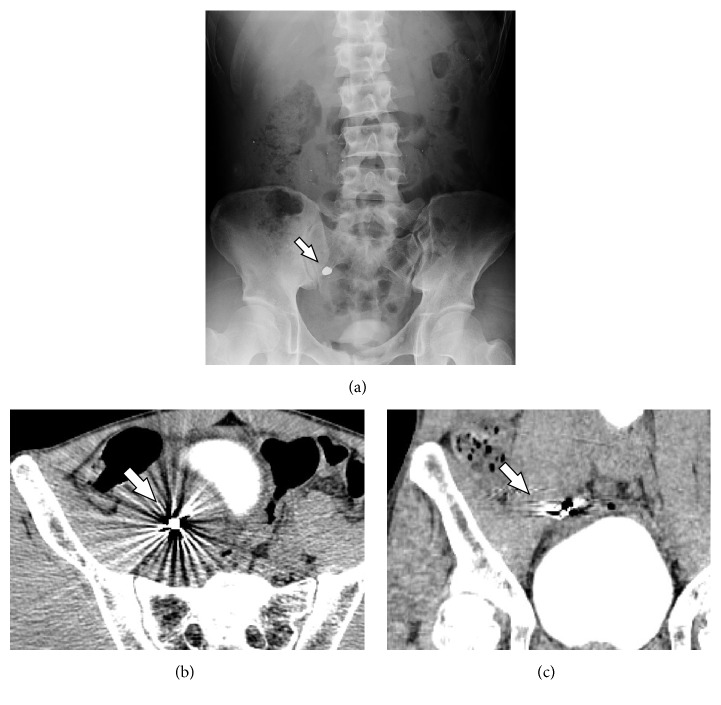
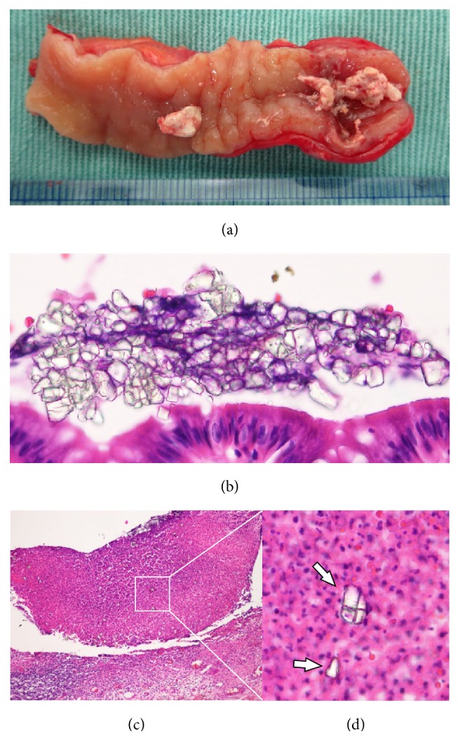
A 41-year-old man presented to our hospital with lower abdominal pain and a high-grade fever. Physical examination revealed rebound tenderness and guarding in the lower abdomen. Abdominal X-ray examination showed a radiopaque object in the right lower quadrant of the abdomen. Abdominal computed tomography (CT) demonstrated that the object had a strong artifact with over 10,000 Hounsfield units, as well as ascites around the terminal ileum. We diagnosed acute peritonitis with a suspicion of the perforation due to unknown foreign body and performed an emergency laparotomy. Operative findings showed a contained perforation of a phlegmonous appendicitis, and appendectomy was performed. The resected specimen demonstrated that the appendix contained a fecalith, and histopathological examination showed the crystal structure of barium sulfate in the lumen of the appendix. Unfortunately, we did not obtain the history of screening for gastric cancer using a barium examination one month prior to our appendectomy. Our experience demonstrates the importance of establishing a history of barium examinations of the gastrointestinal tract in a patient with a radiopaque object in the right lower quadrant of the abdomen for early diagnosis of barium appendicitis. Additionally, early diagnosis of barium appendicitis may affect the selection of surgical procedures.
Figures
References
Publication types
LinkOut - more resources
Full Text Sources
Other Literature Sources
