

Sequence and Timing of Intracranial Changes in Cytomegalovirus in Pregnancy: A Case Report and Literature Review
- PMID: 28487795
- PMCID: PMC5402237
- DOI: 10.1155/2017/5928398
Sequence and Timing of Intracranial Changes in Cytomegalovirus in Pregnancy: A Case Report and Literature Review
Abstract
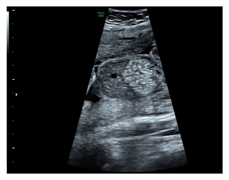
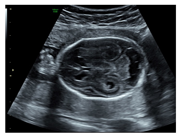
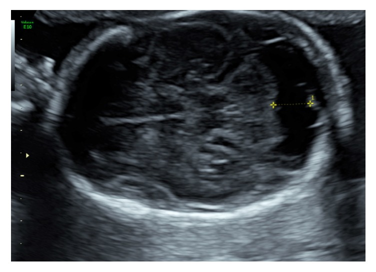
Cytomegalovirus (CMV) is the most common cause of intrauterine infection, occurring in up to 2% of all live births. Most women are asymptomatic or experience nonspecific symptoms, which can lead to long-term sequelae in newborns including neurological impairment, hearing loss, and mental retardation. A 41-year-old woman (G6 P2), with a medical history of epilepsy, presented for her routine anomaly scan at 20 + 4/40. A single finding of echogenic bowel was noted on ultrasound which prompted a full investigation. A repeat ultrasound only five days later demonstrated progressive changes, which included bilateral ventriculomegaly with oedema of the posterior ventricular wall, periventricular hyperechogenicity, and enlargement of the cisterna magna. CMV DNA was detected at amniocentesis. Ultrasound findings are not diagnostic for CMV with only 11-15% of at-risk fetuses being identified. Unfortunately, these findings may be the only indication of an abnormality. There is a well-documented lack of awareness surrounding CMV and screening is not routinely offered. Given the risk to the pregnancy of CMV and to subsequent pregnancies, simple education at the start of a pregnancy could significantly reduce the incidence of maternal CMV.
Figures
Similar articles
-
Prevention and treatment of fetal cytomegalovirus infection with cytomegalovirus hyperimmune globulin: a multicenter study in Madrid.J Matern Fetal Neonatal Med. 2019 Feb;32(4):617-625. doi: 10.1080/14767058.2017.1387890. Epub 2017 Oct 26. J Matern Fetal Neonatal Med. 2019. PMID: 28978246
-
Intracranial ultrasound abnormalities and fetal cytomegalovirus infection: report of 8 cases and review of the literature.Fetal Diagn Ther. 2011;30(2):141-9. doi: 10.1159/000330636. Epub 2011 Sep 29. Fetal Diagn Ther. 2011. PMID: 21952353 Review.
-
Cytomegalovirus infection in pregnancy.J Obstet Gynaecol Can. 2010 Apr;32(4):348-354. doi: 10.1016/S1701-2163(16)34480-2. J Obstet Gynaecol Can. 2010. PMID: 20500943
-
Congenital cytomegalovirus infection in twin pregnancies: viral load in the amniotic fluid and pregnancy outcome.Pediatrics. 2003 Aug;112(2):e153-7. doi: 10.1542/peds.112.2.e153. Pediatrics. 2003. PMID: 12897321
-
Congenital cytomegalovirus infection in pregnancy: a review of prevalence, clinical features, diagnosis and prevention.Aust N Z J Obstet Gynaecol. 2016 Feb;56(1):9-18. doi: 10.1111/ajo.12408. Epub 2015 Sep 22. Aust N Z J Obstet Gynaecol. 2016. PMID: 26391432 Review.
Cited by
-
Diagnostic challenges in congenital cytomegalovirus infection in pregnancy: A case report.Case Rep Womens Health. 2019 Apr 26;22:e00119. doi: 10.1016/j.crwh.2019.e00119. eCollection 2019 Apr. Case Rep Womens Health. 2019. PMID: 31192993 Free PMC article.
-
Ultrasonographic Signs of Cytomegalovirus Infection in the Fetus-A Systematic Review of the Literature.Diagnostics (Basel). 2023 Jul 18;13(14):2397. doi: 10.3390/diagnostics13142397. Diagnostics (Basel). 2023. PMID: 37510141 Free PMC article. Review.
References
-
- Romanelli R. M., Magny J. F., Jacquemard F. Prognostic markers of symptomatic congenital cytomegalovirus infection. Brazilian Journal of Infectious Diseases. 2008;12(1):38–43. - PubMed
Publication types
LinkOut - more resources
Full Text Sources
Other Literature Sources
Research Materials
Miscellaneous
