

Echographic Evaluation of a Subconjunctival Cystic Lesion
- PMID: 28487797
- PMCID: PMC5405365
- DOI: 10.1155/2017/5401850
Echographic Evaluation of a Subconjunctival Cystic Lesion
Abstract
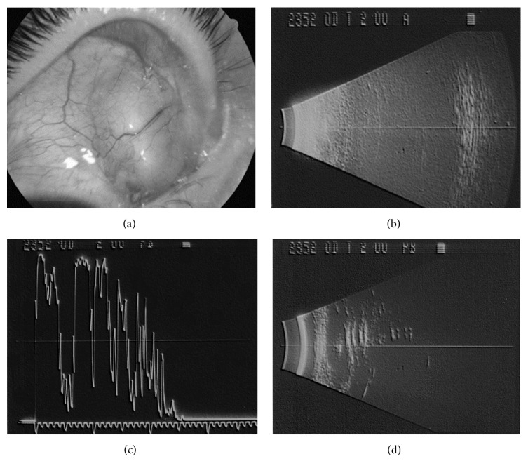
Migration of intraocular silicone oil, used in the treatment of complicated retinal detachment, has been rarely described, but when it happens it can arise with a differential diagnosis with scleral buckling extrusion, tumor, dermoid, ocular cysticercosis, and abscess. The presence of silicone oil in the eye gives very ugly echographic pictures, but these kinds of pictures can be very useful in making a differential diagnosis in the above-mentioned cases. A 39-year-old white female complained of the presence of conjunctival hyperemia and tearing in the right eye (RE); her visual acuity was hand motion, and the intraocular pressure was 14 mmHg. In the upper nasal quadrant a dome shaped lesion was detected. Due to the lens opacities, the patient underwent an echographic examination, which revealed the presence of silicon oil both in the vitreous chamber and in a large subconjunctival space, corresponding to the lesion. This article in addition provides a possible explanation of such cystic formation and discusses the risk factors and the role of the echographic examination in such cases.
Figures
Similar articles
-
Bilateral Asymmetric Rhegmatogenous Retinal Detachment in a Patient with Stickler Syndrome.Turk J Ophthalmol. 2018 Apr;48(2):95-98. doi: 10.4274/tjo.60430. Epub 2018 Apr 25. Turk J Ophthalmol. 2018. PMID: 29755825 Free PMC article.
-
Supplemental scleral buckling for inferior retinal detachment in silicone oil-filled eyes.Retina. 2014 Jun;34(6):1076-82. doi: 10.1097/IAE.0000000000000037. Retina. 2014. PMID: 24240555
-
Scleral fixation intraocular lens as a barrier for silicone oil in traumatic aphakia, aniridia, and recurrent retinal detachment:case report.Retin Cases Brief Rep. 2011 Summer;5(3):256-8. doi: 10.1097/ICB.0b013e3181f047ca. Retin Cases Brief Rep. 2011. PMID: 25390179
-
Management of glaucoma after retinal detachment surgery.Curr Opin Ophthalmol. 2002 Apr;13(2):103-9. doi: 10.1097/00055735-200204000-00009. Curr Opin Ophthalmol. 2002. PMID: 11880724 Review.
-
Lymphatic vessels of the eye - old questions - new insights.Ann Anat. 2019 Jan;221:1-16. doi: 10.1016/j.aanat.2018.08.004. Epub 2018 Sep 18. Ann Anat. 2019. PMID: 30240907 Review.
Cited by
-
Subconjunctival silicone oil complicating strabismus surgery.GMS Ophthalmol Cases. 2019 Apr 4;9:Doc12. doi: 10.3205/oc000101. eCollection 2019. GMS Ophthalmol Cases. 2019. PMID: 31157154 Free PMC article.
-
Ocular ultrasound evaluation of optic nerve sheath diameter in military environments.Mil Med Res. 2019 May 25;6(1):16. doi: 10.1186/s40779-019-0207-8. Mil Med Res. 2019. PMID: 31126318 Free PMC article.
-
Optic nerve ultrasonography to predict increased intracranial pressure in idiopathic intracranial hypertension.Neuroradiol J. 2019 Jun;32(3):227-228. doi: 10.1177/1971400919832001. Epub 2019 Feb 22. Neuroradiol J. 2019. PMID: 30793659 Free PMC article. No abstract available.
References
-
- Biswas J., Bhende P. S., Gopal L., Parikh S., Badrinath S. S. Subconjunctival cysts following silicone oil injection: A clinicopathological study of five cases. Indian Journal of Ophthalmology. 1999;47(3):177–180. - PubMed
Publication types
LinkOut - more resources
Full Text Sources
Other Literature Sources
