Case Reports
. 2017:2017:4097973.
doi: 10.1155/2017/4097973. Epub 2017 Apr 9.

Clinical Challenges in the Diagnosis and Treatment of Temporal Bone Osteomyelitis

Affiliations
Case Reports

Clinical Challenges in the Diagnosis and Treatment of Temporal Bone Osteomyelitis

Liubov Kornilenko et al. Case Rep Otolaryngol. 2017.

Abstract

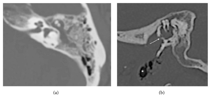
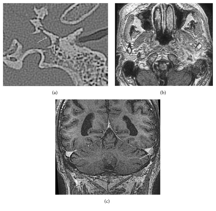
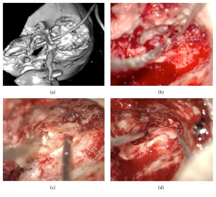
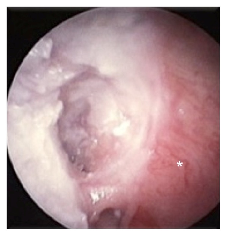
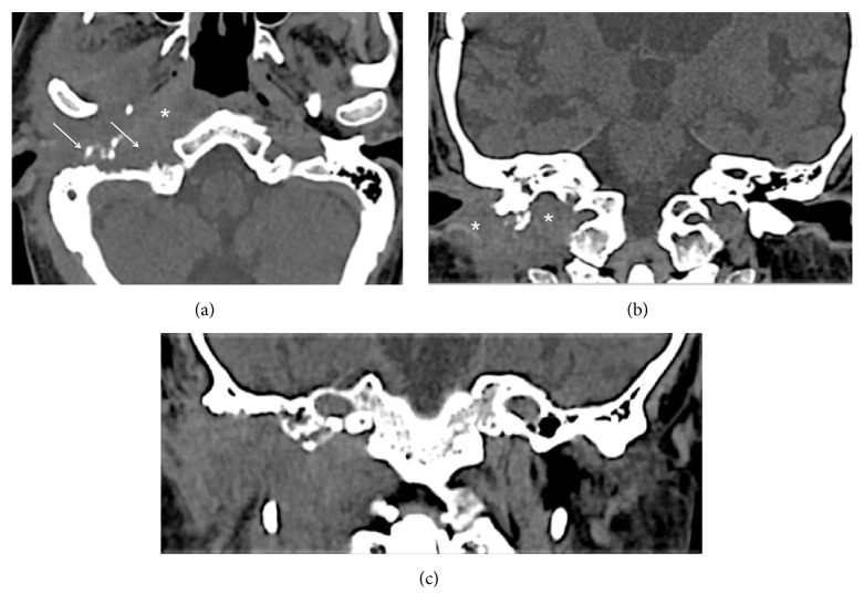
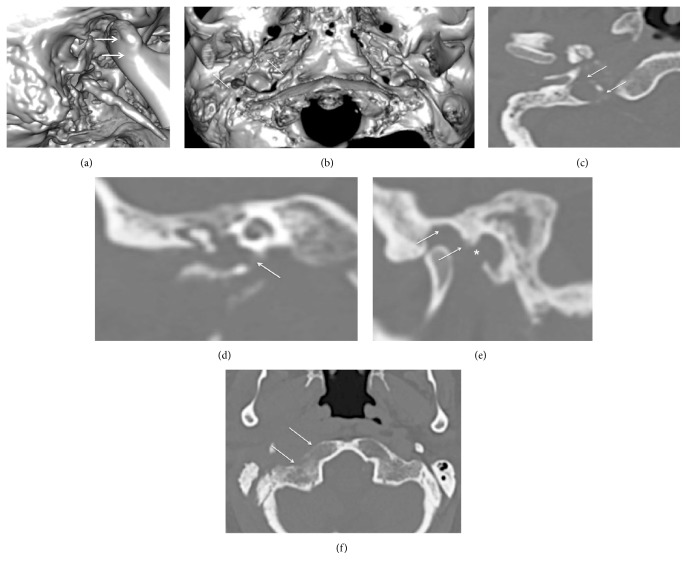
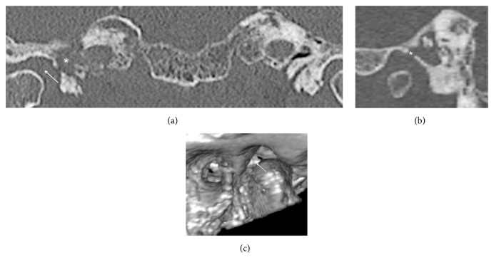
Temporal bone osteomyelitis is a serious life-threatening condition-a quick and proper diagnosis is needed to start treatment and reduce morbidity and mortality. Changing trends of the disease make a differential diagnosis difficult. To emphasize the importance of a clinical suspicion of this dangerous condition, our experience with three difficult cases is presented. The diagnosis was based on clinical symptoms, otoscopic findings, and findings on computed tomography or magnetic resonance imaging. Neoplasm and granulomatous inflammation were excluded by multiple biopsies. The disease can develop in nondiabetic patients. The disorder might be related to the initial inflammatory process in the middle ear with further direct spreading of infection through defects in the bony walls to deep temporal bone structures. Imaging should be performed early to detect osteolytic lesions of the skull base. Surgery was used for the presence of bone sequestra and infratemporal abscess.


Figures

Figure 1
Figure 1
(a) Mastoiditis on the left side. There is diffuse debris throughout the mastoid. (b) An erosion of the anterior-inferior wall of the middle ear cavity (arrow) with air bubbles in the left prestyloid compartment (asterisk).
Figure 2
Figure 2
(a) CT scan of the left temporal bone—a bony sequestrum in the tympanic part is shown by the arrow. (b) An abscess in the left infratemporal fossa appears as a contrast-enhanced mass on the periphery, with irregular contours (arrows). A trismus was explained by inflammatory infiltration of the lateral pterygoid muscles (asterisk). (c) A thrombosed sigmoid sinus is seen on the left side (arrow).
Figure 3
Figure 3
(a) A 3D volume rendering scan shows the amount of bone removed during lateral petrosectomy. The drain was inserted into the abscess cavity through the defect in the anterior EAC wall (arrow). (b) Intraoperative image: a defect in the anterior wall of the tympanic cavity (arrow). Pus under the pressure appeared when the granulations were removed from the tympanic cavity. (c) After the tympanic part of the temporal bone was partially drilled, a large bony sequestrum was removed with microforceps. (d) Finally, the third type of tympanoplasty was carried out.
Figure 4
Figure 4
Six months postoperatively, a partially epithelized mastoid bowl (asterisk) with an intact tympanic membrane was seen.
Figure 5
Figure 5
(a) Inflammatory infiltration at the level of the prevertebral and superior pharyngeal constrictor muscles (asterisk). Arrows show the massive erosion of the tympanic part of the temporal bone. (b) Inflammatory changes at the level of the jugular fossa and parotid compartment (asterisks). (c) A soft tissue lesion in the parapharyngeal and parotid spaces.
Figure 6
Figure 6
(a) The 3D volume rendering technique shows the lateral surface of the temporal bone: the destruction of the base of the styloid process (vaginal process) is clearly seen. Anterior dislocation of the mandible with subluxation of the condyle is shown with arrows. Note slight cortical erosion of the mastoid process of the right side. (b) Inferior view of the skull base: massive destruction of the petrous and tympanic parts of the temporal bone is marked with arrows. (c) The most severe bone lytic changes were observed around the carotid foramen (arrows). (d) A destruction of the carotid plate is clearly seen on this coronal reformat (arrow). (e) The other possible infection-spreading pathway was from the external auditory canal. Asterisk shows a defect in the anterior wall of the latter. Additionally, arrows show erosion of the mandibular fossa roof as well as auricular tubercle. Note an anterior dislocation of the mandibular condyle. (f) Axial CT scan shows the destruction of the cortical layer of the occipital bone (arrows).
Figure 7
Figure 7
(a) The asterisk shows erosion of the temporal bone around the carotid canal (peritubal mastoiditis). The arrow shows a widened TMJ cavity. (b) Defect in the anterior tympanic cavity wall (asterisk). (c) A 3D volume rendering technique shows the same defect from the fossa mandibularis perspective (arrow).

Similar articles

Cited by

References

    1. Thio D., Reece P., Herdman R. Necrotizing otitis externa: a painless reminder. European Archives of Oto-Rhino-Laryngology. 2008;265(8):907–910. doi: 10.1007/s00405-008-0696-7. - DOI - PubMed
    1. Slattery W. H., III, Brackmann D. E. Skull base osteomyelitis: malignant external otitis. Otolaryngologic Clinics of North America. 1996;29(5):795–806. - PubMed
    1. Kim C.-W., Lee J. J., Kim H.-S., Won J.-Y. Skull base osteomyelitis following a mastoidectomy. Auris Nasus Larynx. 2013;40(6):584–586. doi: 10.1016/j.anl.2012.12.002. - DOI - PubMed
    1. Merchant S., Vernick D. M. Osteomyelitis of the temporal bone and skull base in diabetes resulting from otitis media. Skull Base. 1992;2(4):207–212. doi: 10.1055/s-2008-1057137. - DOI - PMC - PubMed
    1. Dudkiewicz M., Livni G., Kornreich L., Nageris B., Ulanovski D., Raveh E. Acute mastoiditis and osteomyelitis of the temporal bone. International Journal of Pediatric Otorhinolaryngology. 2005;69(10):1399–1405. doi: 10.1016/j.ijporl.2005.03.036. - DOI - PubMed

Publication types

LinkOut - more resources