

Transcriptional and Molecular Pathways Activated in Mesenteric Adipose Tissue and Intestinal Mucosa of Crohn's Disease Patients
- PMID: 28487813
- PMCID: PMC5401739
- DOI: 10.1155/2017/7646859
Transcriptional and Molecular Pathways Activated in Mesenteric Adipose Tissue and Intestinal Mucosa of Crohn's Disease Patients
Abstract
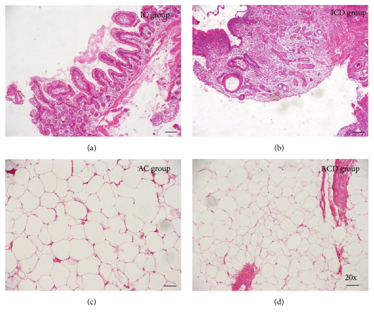
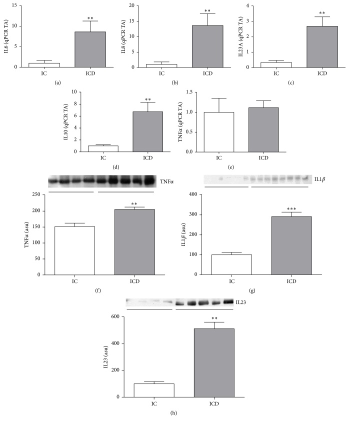
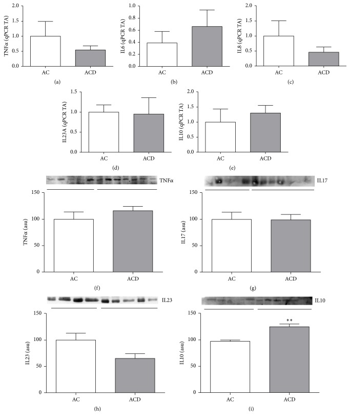
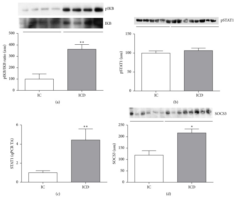
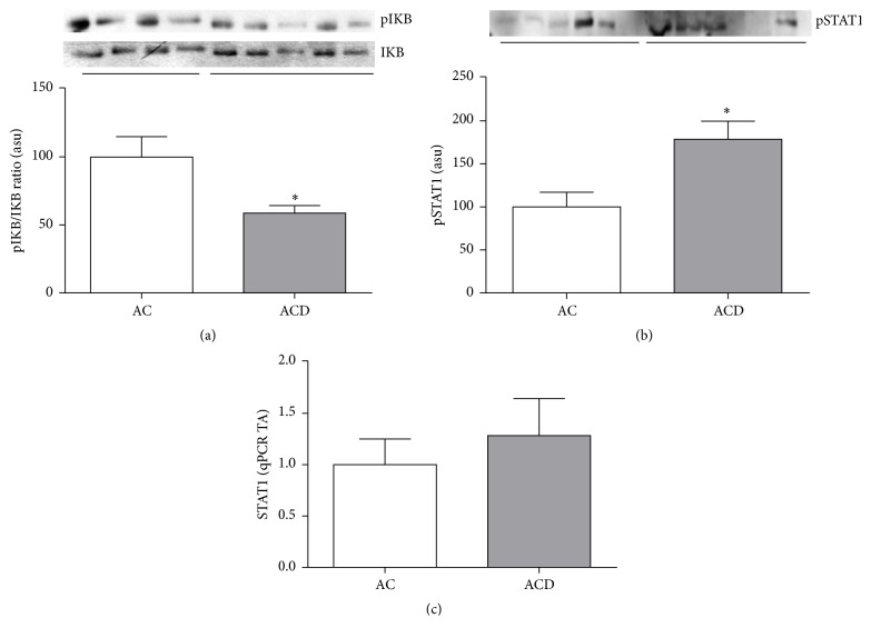
Crohn's disease (CD) is a chronic inflammatory disorder, characterized by cytokine imbalance and transcription signaling pathways activation. In addition, the increase of mesenteric adipose tissue (MAT) near the affected intestinal area is a hallmark of CD. Therefore, we evaluated the transcription signaling pathways and cytokines expression in intestinal mucosa and MAT of active CD patients. Ten patients with ileocecal CD and eight with noninflammatory diseases were studied. The biopsies of intestinal mucosa and MAT were snap-frozen and protein expression was determined by immunoblotting. RNA levels were measured by qPCR. The pIkB/IkB ratio and TNFα level were significantly higher in intestinal mucosa of CD when compared to controls. However, STAT1 expression was similar between intestinal mucosa of CD and controls. Considering the MAT, the pIkB/IkB ratio was significantly lower and the anti-inflammatory cytokine IL10 was significantly higher in CD when compared to controls. Finally, the protein content of pSTAT1 was higher in MAT of CD compared to controls. These findings reinforce the predominance of the proinflammatory NF-kB pathway in CD intestinal mucosa. For the first time, we showed the activation of STAT1 pathway in MAT of CD patients, which may help to understand the physiopathology of this immune mediated disease.
Figures
Similar articles
-
Whole transcriptional analysis identifies markers of B, T and plasma cell signaling pathways in the mesenteric adipose tissue associated with Crohn's disease.J Transl Med. 2020 Jan 30;18(1):44. doi: 10.1186/s12967-020-02220-3. J Transl Med. 2020. PMID: 32000799 Free PMC article.
-
Defective apoptosis in intestinal and mesenteric adipose tissue of Crohn's disease patients.PLoS One. 2014 Jun 2;9(6):e98547. doi: 10.1371/journal.pone.0098547. eCollection 2014. PLoS One. 2014. PMID: 24887376 Free PMC article.
-
Toll-like receptor 4, F4/80 and pro-inflammatory cytokines in intestinal and mesenteric fat tissue of Crohn's disease.Int J Clin Exp Med. 2013;6(2):98-104. Epub 2013 Jan 26. Int J Clin Exp Med. 2013. PMID: 23386912 Free PMC article.
-
Medical and Surgical Implications of Mesenteric Adipose Tissue in Crohn's Disease: A Review of the Literature.Inflamm Bowel Dis. 2023 Mar 1;29(3):458-469. doi: 10.1093/ibd/izac120. Inflamm Bowel Dis. 2023. PMID: 35731568 Review.
-
Role of mesenteric component in Crohn's disease: A friend or foe?World J Gastrointest Surg. 2021 Dec 27;13(12):1536-1549. doi: 10.4240/wjgs.v13.i12.1536. World J Gastrointest Surg. 2021. PMID: 35070062 Free PMC article. Review.
Cited by
-
Discovery of a novel RSK2 inhibitor for the treatment of metastatic pancreatic cancer.J Enzyme Inhib Med Chem. 2025 Dec;40(1):2538673. doi: 10.1080/14756366.2025.2538673. Epub 2025 Aug 5. J Enzyme Inhib Med Chem. 2025. PMID: 40762406 Free PMC article.
-
Icariin Inhibits Intestinal Inflammation of DSS-Induced Colitis Mice Through Modulating Intestinal Flora Abundance and Modulating p-p65/p65 Molecule.Turk J Gastroenterol. 2021 Apr;32(4):382-392. doi: 10.5152/tjg.2021.20282. Turk J Gastroenterol. 2021. PMID: 34231485 Free PMC article.
-
Serial Changes in Body Composition and the Association with Disease Activity during Treatment in Patients with Crohn's Disease.Diagnostics (Basel). 2022 Nov 15;12(11):2804. doi: 10.3390/diagnostics12112804. Diagnostics (Basel). 2022. PMID: 36428862 Free PMC article.
-
Whole transcriptional analysis identifies markers of B, T and plasma cell signaling pathways in the mesenteric adipose tissue associated with Crohn's disease.J Transl Med. 2020 Jan 30;18(1):44. doi: 10.1186/s12967-020-02220-3. J Transl Med. 2020. PMID: 32000799 Free PMC article.
-
ER stress activation in the intestinal mucosa but not in mesenteric adipose tissue is associated with inflammation in Crohn's disease patients.PLoS One. 2019 Sep 26;14(9):e0223105. doi: 10.1371/journal.pone.0223105. eCollection 2019. PLoS One. 2019. PMID: 31557250 Free PMC article.
References
LinkOut - more resources
Full Text Sources
Other Literature Sources
Research Materials
Miscellaneous
