Functional organization of the medial temporal lobe memory system following neonatal hippocampal lesion in rhesus monkeys
- PMID: 28488186
- PMCID: PMC6018021
- DOI: 10.1007/s00429-017-1441-z
Functional organization of the medial temporal lobe memory system following neonatal hippocampal lesion in rhesus monkeys
Abstract
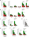
Hippocampal damage in adult humans impairs episodic and semantic memory, whereas hippocampal damage early in life impairs episodic memory but leaves semantic learning relatively preserved. We have previously shown a similar behavioral dissociation in nonhuman primates. Hippocampal lesion in adult monkeys prevents allocentric spatial relational learning, whereas spatial learning persists following neonatal lesion. Here, we quantified the number of cells expressing the immediate-early gene c-fos, a marker of neuronal activity, to characterize the functional organization of the medial temporal lobe memory system following neonatal hippocampal lesion. Ninety minutes before brain collection, three control and four adult monkeys with bilateral neonatal hippocampal lesions explored a novel environment to activate brain structures involved in spatial learning. Three other adult monkeys with neonatal hippocampal lesions remained in their housing quarters. In unlesioned monkeys, we found high levels of c-fos expression in the intermediate and caudal regions of the entorhinal cortex, and in the perirhinal, parahippocampal, and retrosplenial cortices. In lesioned monkeys, spatial exploration induced an increase in c-fos expression in the intermediate field of the entorhinal cortex, the perirhinal, parahippocampal, and retrosplenial cortices, but not in the caudal entorhinal cortex. These findings suggest that different regions of the medial temporal lobe memory system may require different types of interaction with the hippocampus in support of memory. The caudal perirhinal cortex, the parahippocampal cortex, and the retrosplenial cortex may contribute to spatial learning in the absence of functional hippocampal circuits, whereas the caudal entorhinal cortex may require hippocampal output to support spatial learning.
Keywords: Cingulate; Entorhinal; Hippocampus; Parahippocampal; Perirhinal; Retrosplenial.
Figures


Similar articles
-
Severity of memory impairment in monkeys as a function of locus and extent of damage within the medial temporal lobe memory system.Hippocampus. 1994 Aug;4(4):483-95. doi: 10.1002/hipo.450040410. Hippocampus. 1994. PMID: 7874239
-
Damage limited to the hippocampal region produces long-lasting memory impairment in monkeys.J Neurosci. 1995 May;15(5 Pt 2):3796-807. doi: 10.1523/JNEUROSCI.15-05-03796.1995. J Neurosci. 1995. PMID: 7751947 Free PMC article.
-
Lesions of perirhinal and parahippocampal cortex that spare the amygdala and hippocampal formation produce severe memory impairment.J Neurosci. 1989 Dec;9(12):4355-70. doi: 10.1523/JNEUROSCI.09-12-04355.1989. J Neurosci. 1989. PMID: 2593004 Free PMC article.
-
Contrasting hippocampal and perirhinal cortex function using immediate early gene imaging.Q J Exp Psychol B. 2005 Jul-Oct;58(3-4):218-33. doi: 10.1080/02724990444000131. Q J Exp Psychol B. 2005. PMID: 16194966 Review.
-
The medial temporal lobe.Annu Rev Neurosci. 2004;27:279-306. doi: 10.1146/annurev.neuro.27.070203.144130. Annu Rev Neurosci. 2004. PMID: 15217334 Review.
Cited by
-
Structural plasticity in the entorhinal and perirhinal cortices following hippocampal lesions in rhesus monkeys.Hippocampus. 2023 Oct;33(10):1094-1112. doi: 10.1002/hipo.23567. Epub 2023 Jun 19. Hippocampus. 2023. PMID: 37337377 Free PMC article.
-
Life and Death of Immature Neurons in the Juvenile and Adult Primate Amygdala.Int J Mol Sci. 2021 Jun 22;22(13):6691. doi: 10.3390/ijms22136691. Int J Mol Sci. 2021. PMID: 34206571 Free PMC article.
-
Anatomo-functional changes in neural substrates of cognitive memory in developmental amnesia: Insights from automated and manual Magnetic Resonance Imaging examinations.Hippocampus. 2024 Nov;34(11):645-658. doi: 10.1002/hipo.23638. Epub 2024 Sep 13. Hippocampus. 2024. PMID: 39268888
-
Stereological analysis of the rhesus monkey entorhinal cortex.J Comp Neurol. 2018 Sep 1;526(13):2115-2132. doi: 10.1002/cne.24496. Epub 2018 Aug 8. J Comp Neurol. 2018. PMID: 30004581 Free PMC article.
-
Postnatal development of the entorhinal cortex: A stereological study in macaque monkeys.J Comp Neurol. 2020 Oct;528(14):2308-2332. doi: 10.1002/cne.24897. Epub 2020 Mar 18. J Comp Neurol. 2020. PMID: 32134112 Free PMC article.
References
-
- Abrahams S, Pickering A, Polkey CE, Morris RG. Spatial memory deficits in patients with unilateral damage to the right hippocampal formation. Neuropsychologia. 1997;35(1):11–24. - PubMed
-
- Aguirre GK, D’Esposito M. Topographical disorientation: a synthesis and taxonomy. Brain. 1999;122( Pt 9):1613–1628. - PubMed
-
- Albasser MM, Poirier GL, Warburton EC, Aggleton JP. Hippocampal lesions halve immediate-early gene protein counts in retrosplenial cortex: distal dysfunctions in a spatial memory system. Eur J Neurosci. 2007;26(5):1254–1266. - PubMed
-
- Amaral DG, Insausti R, Cowan WM. The entorhinal cortex of the monkey: I. Cytoarchitectonic organization. J Comp Neurol. 1987;264(3):326–355. - PubMed
MeSH terms
Substances
Grants and funding
- R01 NS016980/NS/NINDS NIH HHS/United States
- P51 OD011107/OD/NIH HHS/United States
- 310030_143956/Schweizerischer Nationalfonds zur Förderung der Wissenschaftlichen Forschung
- R37 MH041479/MH/NIMH NIH HHS/United States
- PP00P3-124536/Schweizerischer Nationalfonds zur Förderung der Wissenschaftlichen Forschung
LinkOut - more resources
Full Text Sources
Other Literature Sources
Medical

