

Functional organization of the medial temporal lobe memory system following neonatal hippocampal lesion in rhesus monkeys
- PMID: 28488186
- PMCID: PMC6018021
- DOI: 10.1007/s00429-017-1441-z
Functional organization of the medial temporal lobe memory system following neonatal hippocampal lesion in rhesus monkeys
Abstract
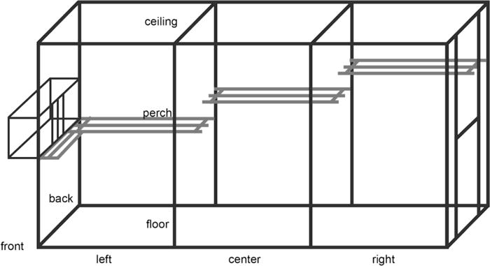
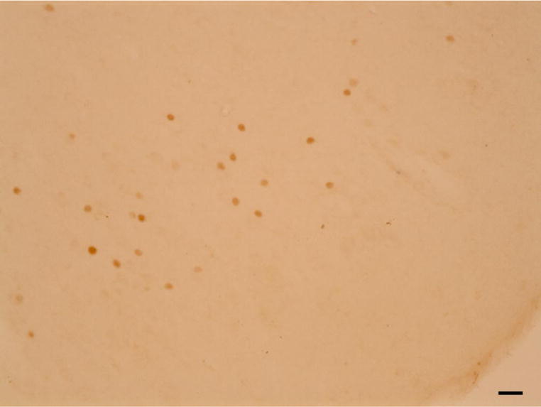
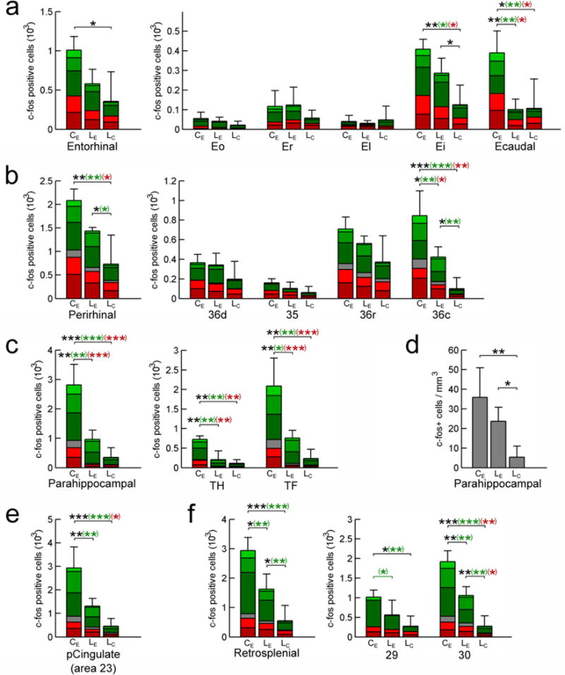
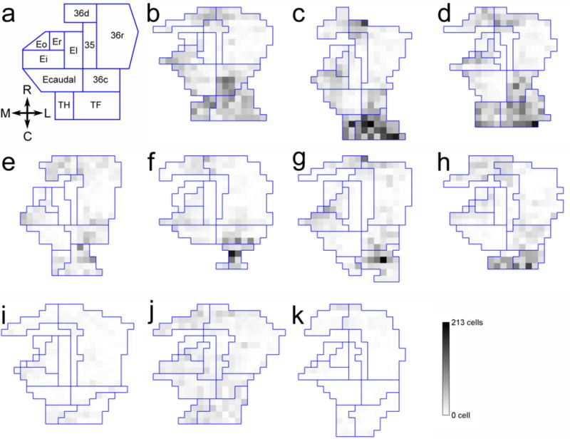
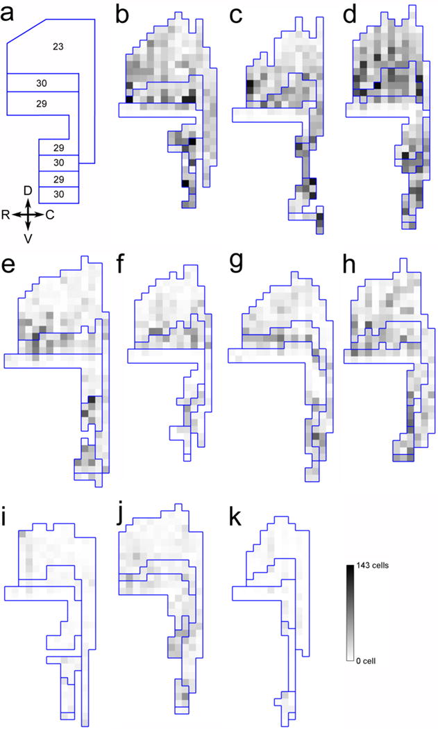
Hippocampal damage in adult humans impairs episodic and semantic memory, whereas hippocampal damage early in life impairs episodic memory but leaves semantic learning relatively preserved. We have previously shown a similar behavioral dissociation in nonhuman primates. Hippocampal lesion in adult monkeys prevents allocentric spatial relational learning, whereas spatial learning persists following neonatal lesion. Here, we quantified the number of cells expressing the immediate-early gene c-fos, a marker of neuronal activity, to characterize the functional organization of the medial temporal lobe memory system following neonatal hippocampal lesion. Ninety minutes before brain collection, three control and four adult monkeys with bilateral neonatal hippocampal lesions explored a novel environment to activate brain structures involved in spatial learning. Three other adult monkeys with neonatal hippocampal lesions remained in their housing quarters. In unlesioned monkeys, we found high levels of c-fos expression in the intermediate and caudal regions of the entorhinal cortex, and in the perirhinal, parahippocampal, and retrosplenial cortices. In lesioned monkeys, spatial exploration induced an increase in c-fos expression in the intermediate field of the entorhinal cortex, the perirhinal, parahippocampal, and retrosplenial cortices, but not in the caudal entorhinal cortex. These findings suggest that different regions of the medial temporal lobe memory system may require different types of interaction with the hippocampus in support of memory. The caudal perirhinal cortex, the parahippocampal cortex, and the retrosplenial cortex may contribute to spatial learning in the absence of functional hippocampal circuits, whereas the caudal entorhinal cortex may require hippocampal output to support spatial learning.
Keywords: Cingulate; Entorhinal; Hippocampus; Parahippocampal; Perirhinal; Retrosplenial.
Figures
References
-
- Abrahams S, Pickering A, Polkey CE, Morris RG. Spatial memory deficits in patients with unilateral damage to the right hippocampal formation. Neuropsychologia. 1997;35(1):11–24. - PubMed
-
- Aguirre GK, D’Esposito M. Topographical disorientation: a synthesis and taxonomy. Brain. 1999;122( Pt 9):1613–1628. - PubMed
-
- Albasser MM, Poirier GL, Warburton EC, Aggleton JP. Hippocampal lesions halve immediate-early gene protein counts in retrosplenial cortex: distal dysfunctions in a spatial memory system. Eur J Neurosci. 2007;26(5):1254–1266. - PubMed
-
- Amaral DG, Insausti R, Cowan WM. The entorhinal cortex of the monkey: I. Cytoarchitectonic organization. J Comp Neurol. 1987;264(3):326–355. - PubMed
MeSH terms
Substances
Grants and funding
- R01 NS016980/NS/NINDS NIH HHS/United States
- P51 OD011107/OD/NIH HHS/United States
- 310030_143956/Schweizerischer Nationalfonds zur Förderung der Wissenschaftlichen Forschung
- R37 MH041479/MH/NIMH NIH HHS/United States
- PP00P3-124536/Schweizerischer Nationalfonds zur Förderung der Wissenschaftlichen Forschung
LinkOut - more resources
Full Text Sources
Other Literature Sources
Medical
