. 2018 Jan;181(1):54-61.
doi: 10.1007/s12011-017-1044-9. Epub 2017 May 9.

Uptake and Utilization of Selenium from Selenoprotein P

Affiliations

Uptake and Utilization of Selenium from Selenoprotein P

Sumangala Shetty et al. Biol Trace Elem Res. 2018 Jan.

Abstract

Selenoprotein P (SELENOP) is a serum glycoprotein that is required for proper selenium distribution in mammals, particularly in supplying selenium to the brain and testes. As the sole mechanism for providing essential selenium to developing spermatozoa, SELENOP metabolism is central to male fertility in all mammals. In addition, this process is important for proper brain function, especially under conditions of limited dietary selenium. Several specific and nonspecific mechanisms for SELENOP uptake in target tissues have been described, but the utilization of SELENOP as a source of selenium for intracellular selenoprotein production has not been systematically characterized. In this report, we examine the process of SELENOP uptake using a robust selenium uptake assay that measures selenium utilization in cells fed 75Se-SELENOP. Using a series of inhibitors and modulators we have identified specific regulators of the process and found that SELENOP must be in an oxidized state for uptake. This assay also demonstrates that SELENOP uptake is not highly sequence specific as the zebrafish protein is recognized and processed by mammalian cells.

Keywords: Oxidation; Selenium; Selenocysteine; Selenoprotein P.


Figures
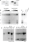
Figure 1
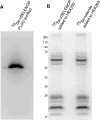
Figure 1. 75Se-SELENOP can be used as a selenium source for intracellular selenoprotein production
(A) Conditioned medium from HepG2 cells grown in the presence of 75Se-selenite was concentrated ~50-fold and a sample was treated with PNGaseF. The samples were resolved by SDS-PAGE and SELENOP was detected by immunoblot (left) and phosphorimage analysis (right). (B) HeLa cells were incubated with ~2 pmol of 75Se-SELENOP or 100 nM 75Se selenite for the time points indicated then switched to normal growth medium and allowed to grow for a total of 24 hours. Endogenous selenoproteins that utilized the 75Se were resolved by SDS-PAGE and detected by PhosphorImage analysis. (C) HeLa cells were labeled with 75Se-SELENOP as described in (A) and lysates were incubated with either an anti-thioredoxin reductase or anti-SELENOP antibody. Immunoprecipitated 75Se-labeled proteins were resolved by SDS-PAGE and detected by PhosphorImage analysis. (D) 75Se-selenite was used to label the endogenous selenoproteins in the cell types indicated: mouse Leydig cells [TM3], mouse Sertoli cells [TM4], human glioblastoma [U87], human hepatoma [HepG2], rat hepatoma [McArdle 7777], human epithelial [HeLa]. After 24 hours of labeling, 4% of the media (left) and 10% of the lysate (right) was resolved by SDS-PAGE and detected by PhosphorImage analysis.
Figure 2
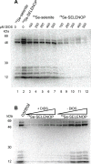
Figure 2. DIDS is a specific inhibitor of SELENOP uptake
(A) HeLa cells were labeled with 75Se selenite or 75Se-SELENOP in the presence of a range of 4,4-Diisothiocyanatostilbene-2,2-Disulfonic Acid (DIDS) as indicated. 75Se labeled proteins were resolved by SDS-PAGE and detected by PhosphorImage analysis. (B) SELENOP was pre-treated with DIDS. Excess DIDS was removed by gel filtration and varying amounts of the DIDS-treated or untreated control protein was used to label endogenous HeLa selenoproteins as above.
Figure 3
Figure 3. Reduced SELENOP cannot deliver selenium to cells
(A) 75Se-SELENOP was incubated with 10 mM DTT for 1h at 37 °C. DTT was removed by gel filtration and the protein was then used to label endogenous HeLa selenoproteins as described in Figure 1A. (B) Reduced SELENOP was incubated with 25 mM iodoacetamide (IA). Excess IA was removed by gel filtration and the protein was used to label endogenous HeLa selenoproteins as above.
Figure 4
Figure 4. Inhibitors reveal mechanistic details about SELENOP uptake and processing
(A) HeLa cells were treated with the indicated concentrations of Auranofin for 1 hour prior to the addition of 75Se-selenite or 75Se-SELENOP. After 24 hours, the cells were lysed and 75Se labeled proteins were resolved by SDS-PAGE and detected by PhosphorImage analysis. (B) (C) HeLa cells were labeled with 75Se selenite or 75Se-SELENOP in the presence or absence of 10% fetal bovine serum as indicated. Note that the relative intensity of endogenous selenoproteins labeled by 75Se-selenite or 75Se-SELENOP varied due to the use of two different batches of 75Se that differed in specific activity.
Figure 5
Figure 5. Degradation of SELENOP occurs under acidic and reducing conditions
(A) HepG2 derived conditioned media containing 75Se-SELENOP was incubated at 37 °C for 30 min in varying concentrations of acetic acid and/or 2 mM DTT as indicated. (B) 75Se-SELENOP was purified by nickel affinity chromatography and incubated at 37 °C for 30 min.
Figure 6
Figure 6. Fish SELENOP can also be taken up and processed
Conditioned medium derived from HEK-293 cells stably expressing C-terminally FLAG tagged zebrafish SELENOP were labeled with equal 75Se-selenite and the labeled protein purified by anti-FLAG immunoaffinity purification. The resulting purified protein was analyzed by SDS-PAGE followed by PhosphorImage analysis. (B) The purified SELENOP shown in (A) was applied to naive HEK-293 cells and lysates were analyzed by SDS-PAGE after 24 hours. The labeling of HEK 293 cells with an equal of 75Se-selenite is shown as a control as indicated.

Similar articles

Cited by

References

    1. Burk RF, Gregory PE. Some characteristics of 75Se-P, a selenoprotein found in rat liver and plasma, and comparison of it with selenoglutathione peroxidase. Arch Biochem Biophys. 1982;213(1):73–80. - PubMed
    1. Motsenbocker MA, Tappel AL. A selenocysteine-containing selenium-transport protein in rat plasma. Biochim Biophys Acta. 1982;719(1):147–153. - PubMed
    1. Motchnik PA, Tappel AL. Multiple selenocysteine content of selenoprotein P in rats. J Inorg Biochem. 1990;40(3):265–269. - PubMed
    1. Hill KE, Lloyd RS, Yang JG, Read R, Burk RF. The cDNA for rat selenoprotein P contains 10 TGA codons in the open reading frame. J Biol Chem. 1991;266(16):10050–10053. - PubMed
    1. Hill KE, Zhou J, McMahan WJ, Motley AK, Atkins JF, Gesteland RF, Burk RF. Deletion of selenoprotein P alters distribution of selenium in the mouse. J Biol Chem. 2003;278(16):13640–13646. - PubMed

LinkOut - more resources