Review
. 2017 Jul;6(7):1620-1630.
doi: 10.1002/sctm.16-0492. Epub 2017 May 10.

Concise Review: Wharton's Jelly: The Rich, but Enigmatic, Source of Mesenchymal Stromal Cells

Affiliations
Review

Concise Review: Wharton's Jelly: The Rich, but Enigmatic, Source of Mesenchymal Stromal Cells

John E Davies et al. Stem Cells Transl Med. 2017 Jul.

Abstract

The umbilical cord has become an increasingly used source of mesenchymal stromal cells for preclinical and, more recently, clinical studies. Despite the increased activity, several aspects of this cell population have been under-appreciated. Key issues are that consensus on the anatomical structures within the cord is lacking, and potentially different populations are identified as arising from a single source. To help address these points, we propose a histologically based nomenclature for cord structures and provide an analysis of their developmental origins and composition. Methods of cell isolation from Wharton's jelly are discussed and the immunophenotypic and clonal characteristics of the cells are evaluated. The perivascular origin of the cells is also addressed. Finally, clinical trials with umbilical cord cells are briefly reviewed. Interpreting the outcomes of the many clinical studies that have been undertaken with mesenchymal stromal cells from different tissue sources has been challenging, for many reasons. It is, therefore, particularly important that as umbilical cord cells are increasingly deployed therapeutically, we strive to better understand the derivation and functional characteristics of the cells from this important tissue source. Stem Cells Translational Medicine 2017;6:1620-1630.

Keywords: Embryology; Mesenchymal stromal cell; Therapy; Wharton's Jelly.


Figures

Figure 1
Figure 1
Registered clinical trials (2009–2016) employing human umbilical cord MSCs numbered a total of 109 as of January 2016, based on Clinicaltrials.gov data, although only 34 are currently open. The pie‐chart shows the broad distribution of target indications (excluding those from cord blood). Although “Haematological” indications are the largest group at 12%, the majority of trials rely on the immune modulatory and anti‐inflammatory properties of the cells, rather than a capacity for connective tissue lineage differentiation. These percentages differ from MSC trials employing cells from all tissue sources, where “Neuro‐degenerative” and “Liver” targets represent 60% of the total number of clinical trials. Abbreviation: MSC, mesenchymal stromal cells.
Figure 2
Figure 2
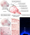
Above: The structure of the human umbilical cord with a three‐dimensional exploded diagram. The diagram was made by directly tracing the outlines of the various features in the histological section, then shifting them along the tilted longitudinal axis. Scale Bar = 5 mm. Below: The human umbilical cord. (A–E): A paraffin embedded section stained with haematoxylin and eosin. (A): Complete cross‐section of the cord showing an outer amniotic epithelium and three vessels contained within Wharton's jelly. The latter is more highly stained in the perivascular zones due to an increase in both cells and matrix. The paucity of staining in those areas beyond the perivascular zones is, in part, due to the presence of clefts [see (B–D)]. Maximum width (a‐a) = 1.8 cm. (B): Enlargement spanning regions 1 and 2 in (A) where the transition from tunica media of the umbilical vein to perivascular Wharton's jelly is clearly seen, as is the transition from perivascular zone to the remainder of Wharton's jelly and the amniotic epithelium. The matrix of the subamniotic Wharton's jelly is marginally denser than that of the intermediate Wharton's jelly, which separates it from the perivascular zone. pv, perivascular zone; sa, subamnion; i, intermediate Wharton's jelly; *, clefts. (C): From area 3 in (A): The amniotic epithelium and the subamniotic Wharton's jelly (sometimes called the “cord lining”). The cells have a stellate morphology. Takechi et al. 27 were the first to report that cells in this subamniotic zone were longer and had more cytoplasmic processes than those in the perivascular zones. *, clefts. (D): From area 2 in (A), the intermediate Wharton's jelly that contains sparse cells, which are small and more rounded in shape, and matrix that surrounds many clefts. *, clefts. (E): From area 1 in (A), the perivascular zone with denser matrix and numerous cells. The latter of varying morphologies, some elongated, some rounded, and others stellate. (F): DAPI (4′,6‐diamidino‐2‐phenylindole) staining of a portion of cord including the vascular tunica media (dense blue structure below), Wharton's Jelly, and the amniotic epithelium (thin dense blue line above). DAPI staining is densest in the perivascular zone and decreases toward the amniotic epithelium. White arrow, perivascular zone. Although the cell density in the amniotic epithelium is clear, there is no obvious increase in cell density in the subamniotic zone compared to that of the iWJ. [Image courtesy of Shiva Hamidian Jahromi]. Scale Bars: A = 5 mm; B = 500 µm; C–E = 100 µm; F = 200 µm. Abbreviation: WJ, Wharton's jelly.
Figure 3
Figure 3
CD146 labeling of a first trimester cord (A) and a term cord (B) counterstained with DAPI. In the first trimester, the smooth muscle vessel wall labels positively, but the surrounding tissue is negative for CD146. On the contrary, at term the vascular endothelium is now highly positive, the vessel wall is still positive, but so also are cells in the perivascular tissue. Scale Bars A, B = 100 µm.
Figure 4
Figure 4
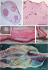
Comparative anatomy of the umbilical cord. (A, B): Porcine, (C‐E): Equine, (F): Canine. (A): Unlike the human cord, the perivascular zones are not clearly demarcated in the porcine cord. (B): In the porcine cord, Wharton's jelly contains many small vascular structures. (C): The equine cord has four major vessels and many co‐lateral and branching vessels. (D): Each of the main vessels has a well‐developed tunica adventitia as seen in this dissected specimen. (E): The tunica adventitia is distinct from the tunica media in this H&E stained cross‐section. (F): Like the equine cord, the canine cord has multiple vessels with co‐lateral branching. The vessels are contained within the amniotic epithelium but, unlike the human, porcine and equine cords, does not from a firm roughly circular but rather a diffuse and flattened structure. [Image courtesy of Dr. Emily Correna Carlo Reis]. Scale Bars: A, B and E = 1 mm; C, D and F = 1 cm.
Figure 5
Figure 5
Due to the high CFU‐F frequency, Sarugaser et al. undertook differentiation assays on both parent and daughter single cell derived clonal populations. Various self‐renewing mesenchymal stromal cells types were identified resulting in the hierarchy show here. The semicircular arrows represent populations that are self‐renewing. The default lineage is the fibroblast. Thus, some stem cells self‐renew and parents and daughters both display five‐lineage differentiation while others are capable of only 4, 3, or 2 lineage differentiation. A self‐renewing fibroblast was also identified. Abbreviations: MACOF, muscle, adipo, chondro, osteo, and fibro; WJ, Wharton's jelly.

Similar articles

Cited by

References

    1. Joerger‐Messerli MS, Marx C, Oppliger B et al. Mesenchymal stem cells from Wharton's jelly and amniotic fluid. Best Pract Res Clin Obstet Gynaecol 2016;31:30–44. - PubMed
    1. Kalaszczynska I, Ferdyn K. Wharton's jelly derived mesenchymal stem cells: Future of regenerative medicine? Recent findings and clinical significance. Biomed Res Int 2015;2015:430847. - PMC - PubMed
    1. Ding D‐C, Chang Y‐H, Shyu W‐C et al. Human umbilical cord mesenchymal stem cells: A new era for stem cell therapy. Cell Transplant 2015;24:339–47. - PubMed
    1. Nagamura‐Inoue T, He H. Umbilical cord‐derived mesenchymal stem cells: Their advantages and potential clinical utility. World J Stem Cells 2014;6:195–202. - PMC - PubMed
    1. El Omar R, Beroud J, Stoltz J‐F et al. Umbilical cord mesenchymal stem cells: The new gold standard for mesenchymal stem cell‐based therapies? Tissue Eng Part B Rev 2014;20:523–544. - PubMed

MeSH terms