

Stimulatory role of interleukin 10 in CD8+ T cells through STATs in gastric cancer
- PMID: 28488547
- PMCID: PMC7221577
- DOI: 10.1177/1010428317706209
Stimulatory role of interleukin 10 in CD8+ T cells through STATs in gastric cancer
Abstract
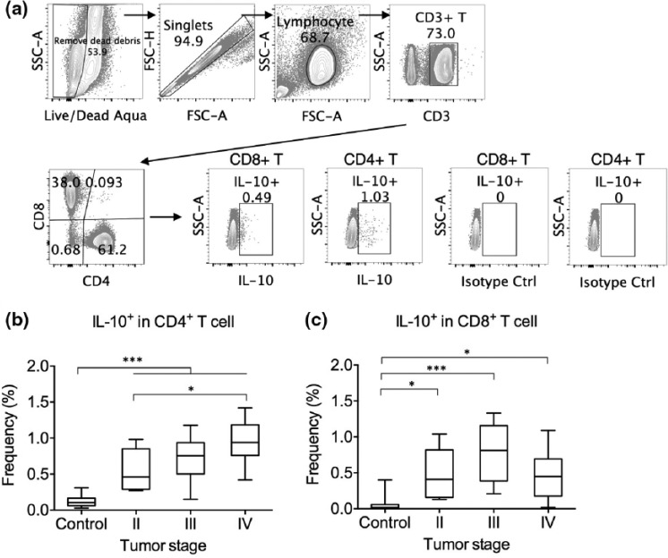
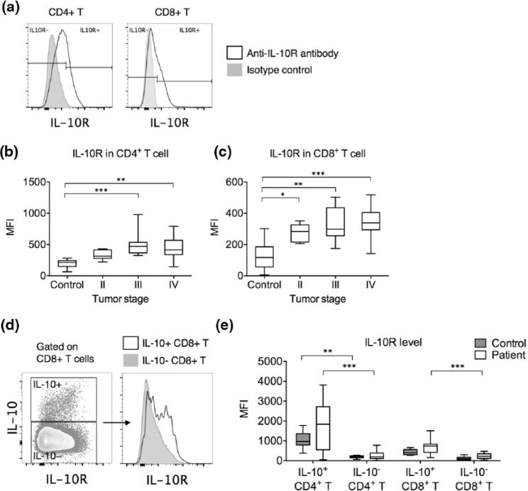
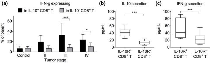
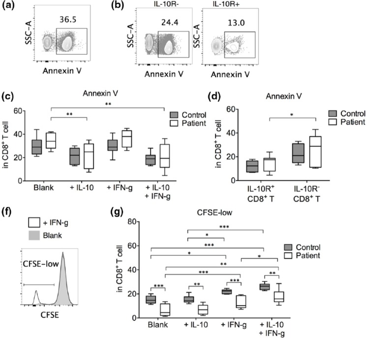
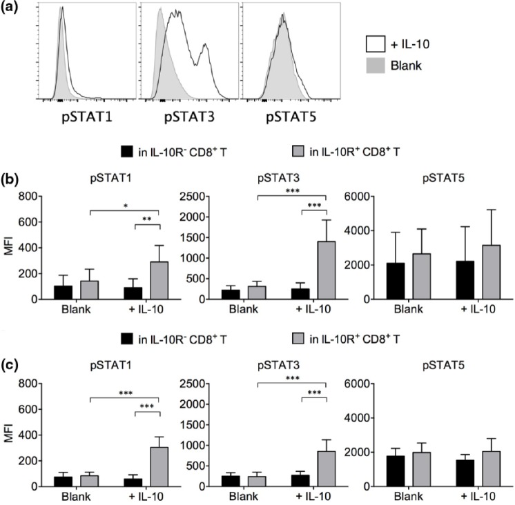
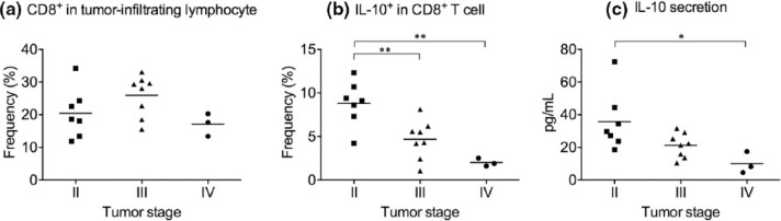
CD8+ T cells are considered to be critical in tumor surveillance and elimination. Increased CD8+ T cell frequency and function is associated with better prognosis in cancer patients. Interleukin 10 is a cytokine with controversial roles in CD8+ T cell-mediated anti-tumor immunity. We therefore examined the interleukin 10 expression and consumption in CD8+ T cells harvested from the peripheral blood and resected tumors of gastric cancer patients of stages II-IV. We found that the gastric cancer patients presented significantly elevated frequencies of interleukin 10-expressing cells in both CD4+ and CD8+ T cells compared to healthy controls. But distinctive from the interleukin 10-expressing CD4+ T cells, which increased in frequency in advanced cancer, the interleukin 10-expressing CD8+ T cells did not increase with cancer stage in the peripheral blood and actually decreased with cancer stage in resected tumor. Interleukin 10 and interleukin 10 receptor expression was also enriched in interferon gamma-expressing activated CD8+ T cells. Compared to interleukin 10-nonexpressing CD8+ T cells, interleukin 10 receptor-expressing CD8+ T cells secreted significantly elevated interferon gamma levels. Treatment of anti-CD3/CD28-stimulated, purified CD8+ T cells with interleukin 10 alone could significantly enhance CD8+ T cell survival, an effect dependent on interleukin 10 receptor expression. Interleukin 10 also increased CD8+ T cell proliferation synergistically with interferon gamma but not alone. Analysis of downstream signal transducer and activator of transcription molecules showed that interleukin 10 treatment significantly increased the phosphorylation of signal transducer and activator of transcription 3 and signal transducer and activator of transcription 1 to lesser extent. Together, these results demonstrate that interleukin 10 possessed stimulatory roles in activated CD8+ T cells from gastric cancer patients.
Keywords: Interleukin 10; gastric cancer.
Conflict of interest statement
Figures
Similar articles
-
Leukotriene B4 receptor 1 is differentially expressed on peripheral T cells of steroid-sensitive and -resistant asthmatics.Ann Allergy Asthma Immunol. 2014 Mar;112(3):211-216.e1. doi: 10.1016/j.anai.2013.12.006. Epub 2014 Jan 7. Ann Allergy Asthma Immunol. 2014. PMID: 24428972 Free PMC article. Clinical Trial.
-
Interleukin-15 differentially enhances the expression of interferon-gamma and interleukin-4 in activated human (CD4+) T lymphocytes.Immunology. 1999 Feb;96(2):207-14. doi: 10.1046/j.1365-2567.1999.00679.x. Immunology. 1999. PMID: 10233697 Free PMC article.
-
IL-15 induces strong but short-lived tumor-infiltrating CD8 T cell responses through the regulation of Tim-3 in breast cancer.Biochem Biophys Res Commun. 2015 Aug 14;464(1):360-6. doi: 10.1016/j.bbrc.2015.06.162. Epub 2015 Jun 30. Biochem Biophys Res Commun. 2015. PMID: 26141233
-
[Interleukin-12 restores and promotes the T-cell immune function inhibited by 5-fluorouracil].Ai Zheng. 2007 Aug;26(8):801-8. Ai Zheng. 2007. PMID: 17697537 Chinese.
-
Increased in vitro induced CD4+ and CD8+ T cell IFN-gamma and CD4+ T cell IL-10 production in stable relapsing multiple sclerosis.Int J Neurosci. 1997 Aug;90(3-4):187-202. doi: 10.3109/00207459709000638. Int J Neurosci. 1997. PMID: 9352427 Review.
Cited by
-
Dual Role of Interleukin-10 in Murine NZB/W F1 Lupus.Int J Mol Sci. 2021 Jan 29;22(3):1347. doi: 10.3390/ijms22031347. Int J Mol Sci. 2021. PMID: 33572870 Free PMC article.
-
Long-term survival after local immunotherapy for malignant gliomas: a retrospective study with 20 years follow-up.BMC Immunol. 2024 Dec 20;25(1):83. doi: 10.1186/s12865-024-00676-2. BMC Immunol. 2024. PMID: 39707189 Free PMC article.
-
The Progress of T Cell Immunity Related to Prognosis in Gastric Cancer.Biomed Res Int. 2018 Feb 27;2018:3201940. doi: 10.1155/2018/3201940. eCollection 2018. Biomed Res Int. 2018. PMID: 29682534 Free PMC article. Review.
-
Comparison of Exosomes Derived from Non- and Gamma-Irradiated Melanoma Cancer Cells as a Potential Antigenic and Immunogenic Source for Dendritic Cell-Based Immunotherapeutic Vaccine.Vaccines (Basel). 2020 Nov 19;8(4):699. doi: 10.3390/vaccines8040699. Vaccines (Basel). 2020. PMID: 33228229 Free PMC article.
-
IL-10-Engineered Dendritic Cells Modulate Allogeneic CD8+ T Cell Responses.Int J Mol Sci. 2023 May 23;24(11):9128. doi: 10.3390/ijms24119128. Int J Mol Sci. 2023. PMID: 37298076 Free PMC article.
References
-
- Ouyang W, Rutz S, Crellin NK, et al. Regulation and functions of the IL-10 family of cytokines in inflammation and disease. Annu Rev Immunol 2011; 29: 71–109. - PubMed
-
- Pestka S, Krause CD, Sarkar D, et al. Interleukin-10 and related cytokines and receptors. Annu Rev Immunol 2004; 22: 929–979. - PubMed
-
- Moore KW, de Waal Malefyt R, Coffman RL, et al. Interleukin-10 and the interleukin-10 receptor. Annu Rev Immunol 2001; 19: 683–765. - PubMed
-
- de Waal Malefyt R, Yssel H, de Vries JE. Direct effects of IL-10 on subsets of human CD4+ T cell clones and resting T cells. Specific inhibition of IL-2 production and proliferation. J Immunol 1993; 150: 4754–4765. - PubMed
-
- Del Prete G, De Carli M, Almerigogna F, et al. Human IL-10 is produced by both type 1 helper (Th1) and type 2 helper (Th2) T cell clones and inhibits their antigen-specific proliferation and cytokine production. J Immunol 1993; 150: 353–360. - PubMed
MeSH terms
Substances
LinkOut - more resources
Full Text Sources
Other Literature Sources
Medical
Research Materials
