. 2017 Nov;232(11):3006-3019.
doi: 10.1002/jcp.25999. Epub 2017 Jun 14.

Hypomorphic conditional deletion of E11/Podoplanin reveals a role in osteocyte dendrite elongation

Affiliations

Hypomorphic conditional deletion of E11/Podoplanin reveals a role in osteocyte dendrite elongation

Katherine A Staines et al. J Cell Physiol. 2017 Nov.

Abstract

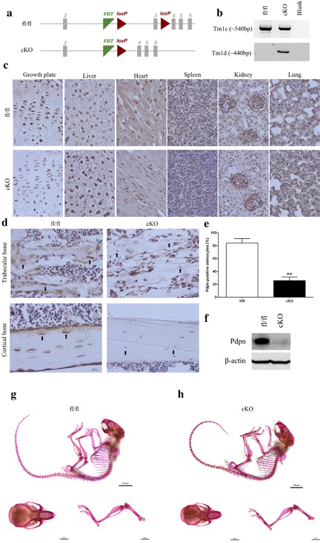
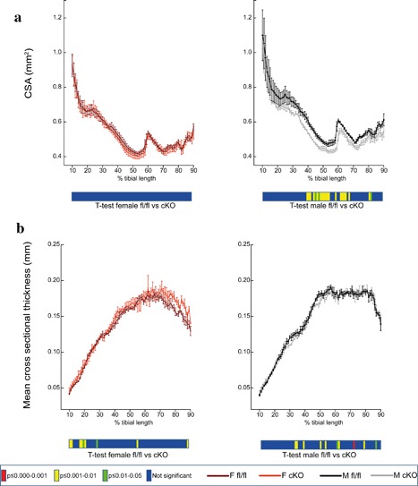
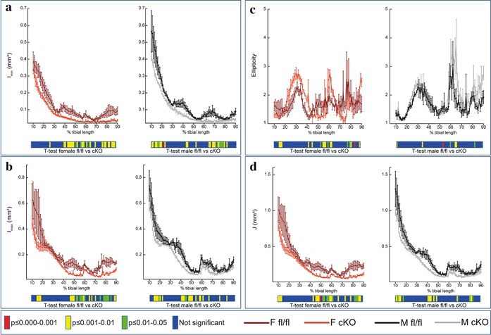
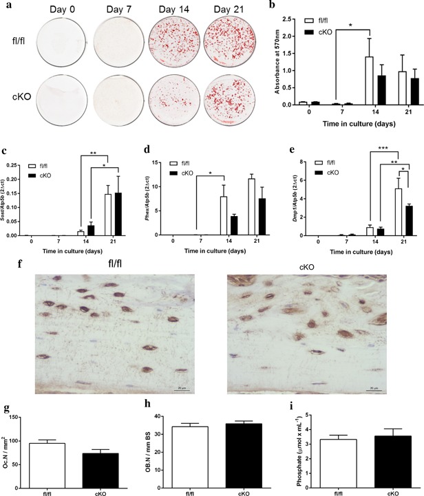
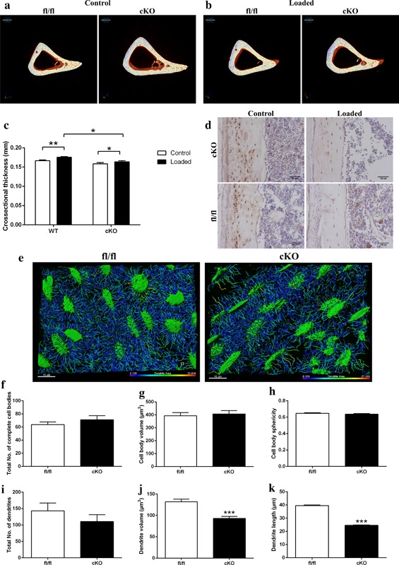
The transmembrane glycoprotein E11/Podoplanin (Pdpn) has been implicated in the initial stages of osteocyte differentiation. However, its precise function and regulatory mechanisms are still unknown. Due to the known embryonic lethality induced by global Pdpn deletion, we have herein explored the effect of bone-specific Pdpn knockdown on osteocyte form and function in the post-natal mouse. Extensive skeletal phenotyping of male and female 6-week-old Oc-cre;Pdpnflox/flox (cKO) mice and their Pdpnflox/flox controls (fl/fl) has revealed that Pdpn deletion significantly compromises tibial cortical bone microarchitecture in both sexes, albeit to different extents (p < 0.05). Consistent with this, we observed an increase in stiffness in female cKO mice in comparison to fl/fl mice (p < 0.01). Moreover, analysis of the osteocyte phenotype by phalloidin staining revealed a significant decrease in the dendrite volume (p < 0.001) and length (p < 0.001) in cKO mice in which deletion of Pdpn also modifies the bone anabolic loading response (p < 0.05) in comparison to age-matched fl/fl mice. Together, these data confirm a regulatory role for Pdpn in osteocyte dendrite formation and as such, in the control of osteocyte function. As the osteocyte dendritic network is known to play vital roles in regulating bone modeling/remodeling, this highlights an essential role for Pdpn in bone homeostasis.

Keywords: E11; dendrite; osteoblasts; osteocalcin; osteocytes.


Figures

Figure 1
Figure 1
(a) Schematic of the Pdpn floxed allele before and after deletion of the loxP cassette containing exon 3 via osteocalcin cre (Oc‐cre) mediated recombination. (b) PCR analysis of genomic DNA from the long bones of fl/fl, and cKO mice with primers for the Tm1c allele (before cre recombination), and the Tm1d allele (∼440 bp, after cre recombination). (c) Immunohistochemical labeling of Pdpn in the lung, kidney, spleen, heart, liver, muscle, and growth plate, of 6‐week‐old mice. Images are representative of n = 4/sex/genotype. Scale bar = 20 μm. (d) Immunohistochemical labeling of Pdpn in the trabecular bone and cortical bone of 6‐week‐old mice. Arrows are pointing at embedded osteocytes within the trabecular and cortical bone and their dendritic processes projecting from the cell bodies. Images are representative of n = 4/sex/genotype. Scale bar = 20μm. (e) Quantification of osteocytes positive for Pdpn immunolabeling relative to negatively labeled osteocytes (n = 3/genotype), p < 0.001***. (f) Western blotting for Pdpn (∼37 kDa) in cortical bone protein lysates from 6‐week‐old mice. β‐actin was used as a loading control. Whole mount Alcian Blue and Alizarin Red stained skeletal preparations of 6‐week‐old male (g) fl/fl, and (h) cKO mice (scale bar = 10 mm) including hindlimb and calvaria preparations (scale bar = 5 mm)
Figure 2
Figure 2
Whole bone analyses of cortical bone between 10% and 90% of total tibial length, excluding proximal and distal metaphyseal bone, of female fl/fl (brown), female cKO (orange), male fl/fl (black), and male cKO (gray) tibia at 6 weeks of age showing (a) cross‐sectional area (CSA; mm2) and (b) mean cross‐sectional thickness (mm). Graphs represent mean ± SEM, n = 4/group. p < 0.05 was considered to be significant and p ≤ 0.01–0.05 was noted as green, p ≤ 0.001–0.01 as yellow, and p ≤ 0.000–0.001 as red. Not significant is noted as blue
Figure 3
Figure 3
Whole bone analyses of cortical bone between 10% and 90% of total tibial length, excluding proximal and distal metaphyseal bone, of female fl/fl (brown), female cKO (orange), male fl/fl (black), and male cKO (gray) tibia at 6 weeks of age showing (a) Imin (mm4), (b) Imax (mm4), (c) ellipticity, and (d) resistance to torsion (J; mm4) Graphs represent mean ± SEM, n = 4/group. p < 0.05 was considered to be significant and p ≤ 0.01–0.05 was noted as green, p ≤ 0.001–0.01 as yellow, and p ≤ 0.000–0.001 as red. Not significant is noted as blue
Figure 4
Figure 4
(a) Alizarin red staining of primary osteoblast cultures from Pdpn cKO and fl/fl mice over a 21‐day culture period. (b) Quantification of Alizarin red staining. RT‐qPCR analysis of osteocyte marker genes (c) Sost, (d) Phex, and (e) Dmp1 in mineralizing primary osteoblast cultures from Pdpn cKO and fl/fl mice over a 21‐day culture period. (f) Immunohistochemical labeling of sclerostin in the cortical bone of 6‐week‐old mice. Arrows are pointing at embedded osteocytes within the cortical bone and their dendritic processes projecting from the cell bodies. Images are representative of n = 4/sex/genotype. Scale bar = 20 μm. (g) Osteoclast cell numbers (OC.N/mm2) in the trabecular bone of 6‐week‐old cKO and fl/fl mice (n = 4/genotype). (h) Osteoblast cell numbers (OB.N/mm bone surface [BS]) in the trabecular bone of 6‐week‐old cKO and fl/fl mice (n = 4/genotype). (i) Serum phosphate levels in cKO and fl/fl mice (n = 6/genotype). Data are represented as mean ± SEM. *p < 0.05, **p < 0.01, ***p < 0.001
Figure 5
Figure 5
Representative images of fl/fl and cKO mice in (a) control and (b) loaded limbs. (c) Cortical cross‐sectional thickness at 37% of the tibia length as assessed by microCT analysis. (d) Immunohistochemical labeling of sclerostin in osteocytes of cortical bone of loaded and control 6‐week‐old Pdpn cKO and fl/fl mice. Arrows are pointing at embedded osteocytes within the cortical bone and their dendritic processes projecting from the cell bodies. Images are representative of n = 4/sex/genotype. Scale bar = 20μm. (e) Laser confocal z‐stack, single channel outlining phalloidin‐Factin staining was imaged in the cortical bone. Imaris cell surface rendering was applied to cell bodies and Imaris FilamentTracer applied to dendritic processes and these were colored according to length. Quantification of osteocyte parameters in 6‐week‐old male fl/fl, and cKO. (f) Total number of complete cell bodies in three volume fields. (g) Cell body volume (μm3). (h) Cell body sphericity. (i) Total number of dendrites in three volume fields. (j) Dendrite volume (μm3). (k) Dendrite length (μm). Data are represented as mean ± SEM. *p < 0.05, **p < 0.01, ***p < 0.001

References

    1. Balemans, W. , Ebeling, M. , Patel, N. , Van Hul, E. , Olson, P. , Dioszegi, M. , … Van Hul, W. (2001). Increased bone density in sclerosteosis is due to the deficiency of a novel secreted protein (SOST). Human Molecular Genetics, 10, 537–543. - PubMed
    1. Cao, Y. X. , Ramirez, M. I. , & Williams, M. C. (2003). Enhanced binding of Sp1/Sp3 transcription factors mediates the hyperoxia‐induced increased expression of the lung type I cell gene T1alpha. Journal of Cellular Biochemistry, 89, 887–901. - PubMed
    1. Dallas, S. L. , Prideaux, M. , & Bonewald, L. F. (2013). The osteocyte: An endocrine cell … and more. Endocrine Reviews, 34, 658–690. - PMC - PubMed
    1. De Souza, R. L. , Matsuura, M. , Eckstein, F. , Rawlinson, S. C. , Lanyon, L. E. , & Pitsillides, A. A. (2005). Non‐invasive axial loading of mouse tibiae increases cortical bone formation and modifies trabecular organization: A new model to study cortical and cancellous compartments in a single loaded element. Bone, 37, 810–818. - PubMed
    1. Dobbs, L. G. , Williams, M. C. , & Gonzalez, R. (1988). Monoclonal antibodies specific to apical surfaces of rat alveolar type I cells bind to surfaces of cultured, but not freshly isolated, type II cells. Biochimica et Biophysica Acta, 970, 146–156. - PubMed