. 2017 May 10:8:15293.
doi: 10.1038/ncomms15293.

Heterodimerization of Munc13 C2A domain with RIM regulates synaptic vesicle docking and priming

Affiliations

Heterodimerization of Munc13 C2A domain with RIM regulates synaptic vesicle docking and priming

Marcial Camacho et al. Nat Commun. .

Abstract

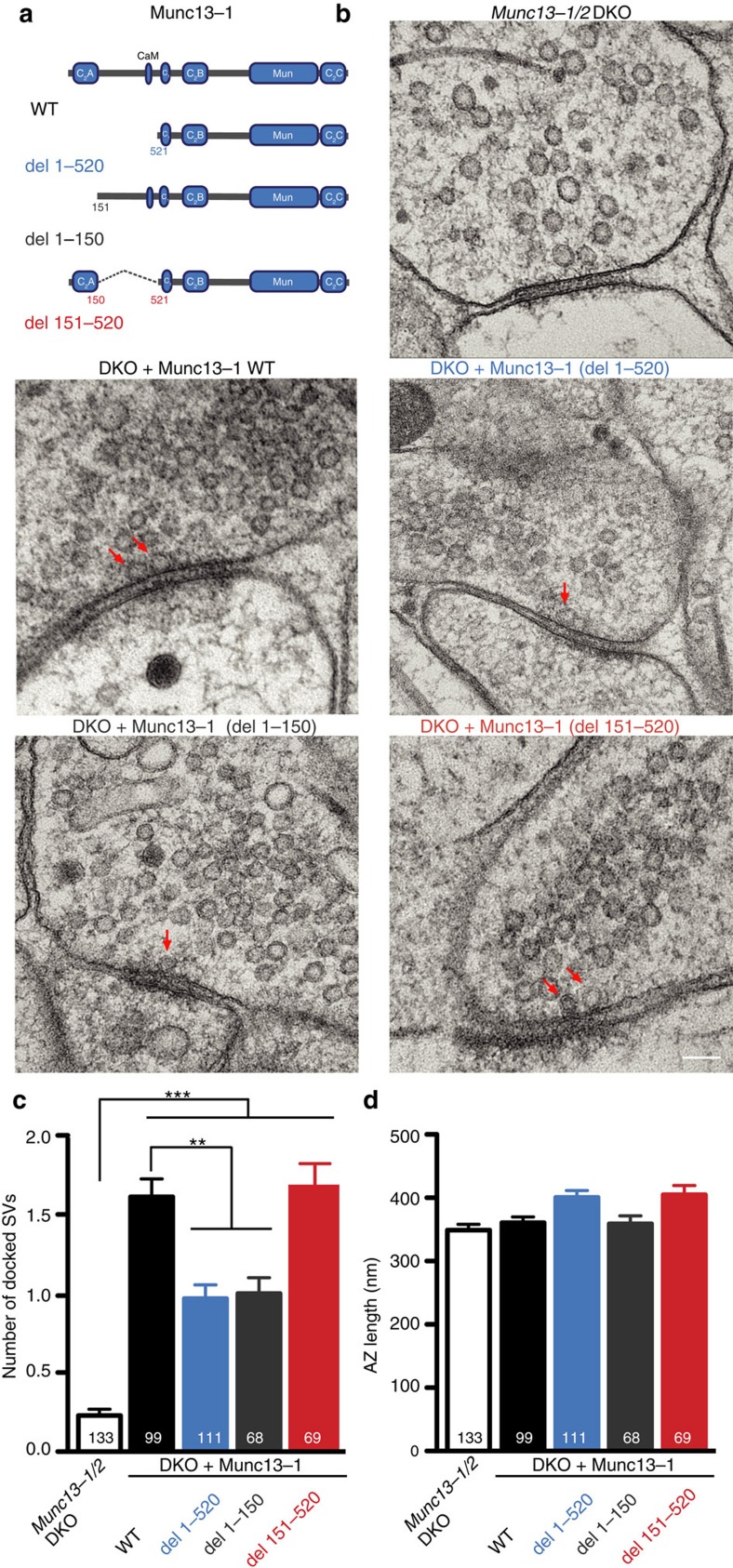
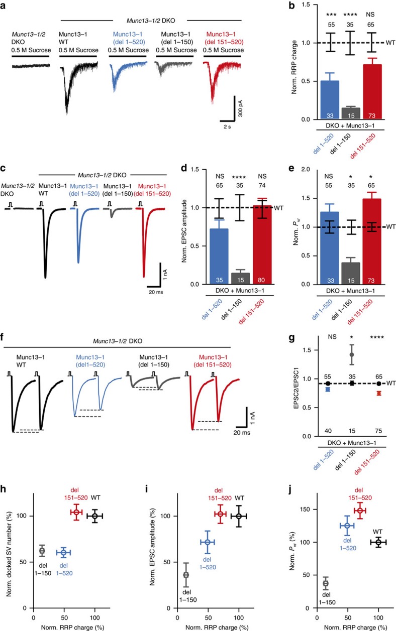
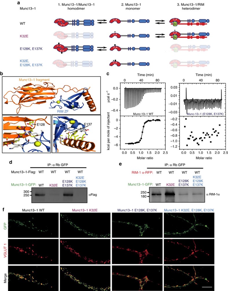
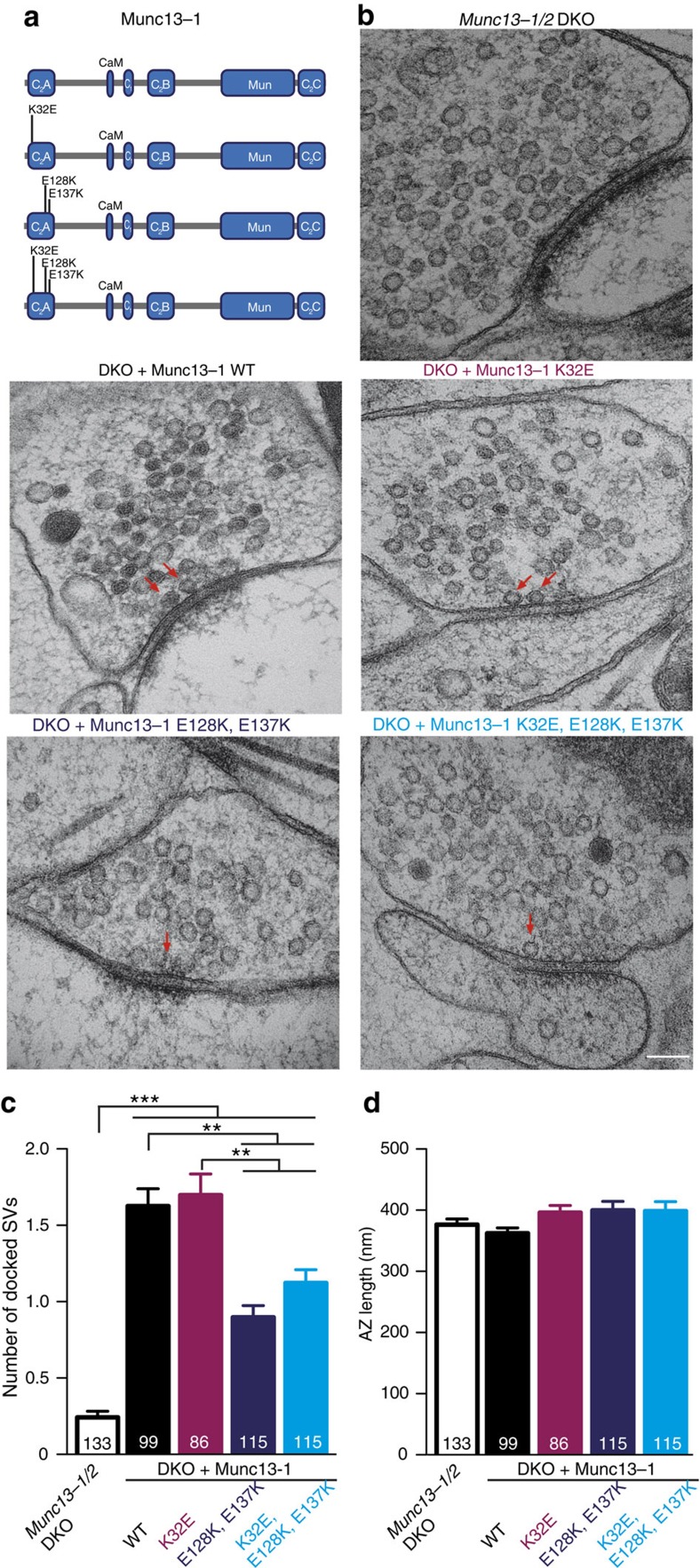
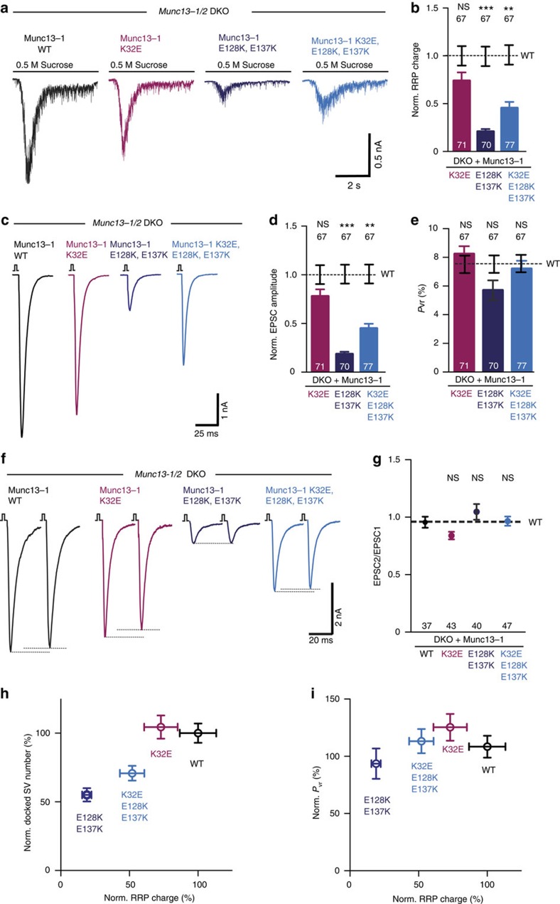
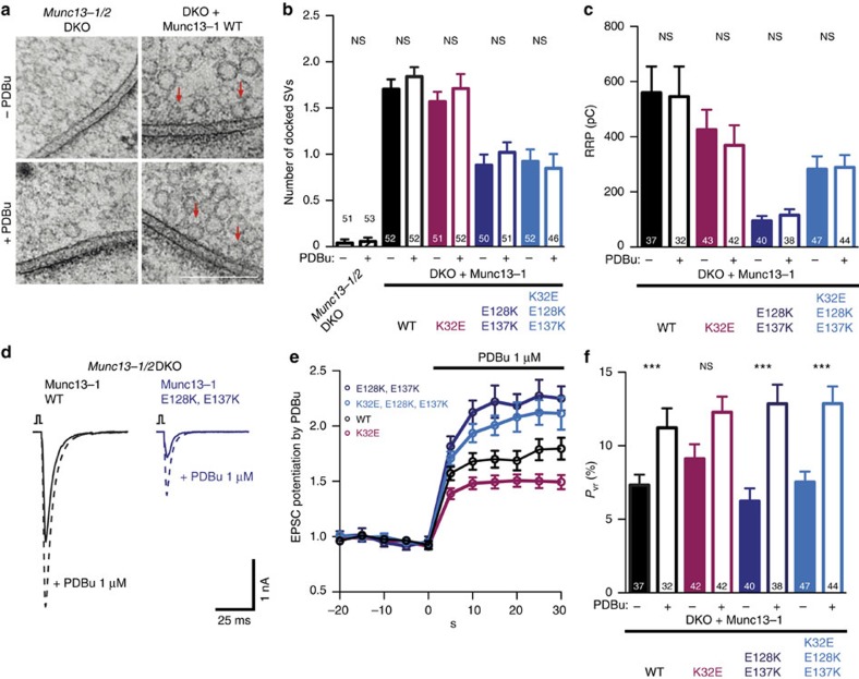
The presynaptic active zone protein Munc13 is essential for neurotransmitter release, playing key roles in vesicle docking and priming. Mechanistically, it is thought that the C2A domain of Munc13 inhibits the priming function by homodimerization, and that RIM disrupts the autoinhibitory homodimerization forming monomeric priming-competent Munc13. However, it is unclear whether the C2A domain mediates other Munc13 functions in addition to this inactivation-activation switch. Here, we utilize mutations that modulate the homodimerization and heterodimerization states to define additional roles of the Munc13 C2A domain. Using electron microscopy and electrophysiology in hippocampal cultures, we show that the C2A domain is critical for additional steps of vesicular release, including vesicle docking. Optimal vesicle docking and priming is only possible when Munc13 heterodimerizes with RIM via its C2A domain. Beyond being a switching module, our data suggest that the Munc13-RIM heterodimer is an active component of the vesicle docking, priming and release complex.


Conflict of interest statement

The authors declare no competing financial interests.

Figures

Figure 1
Figure 1. The N-terminal C2A domain of Munc13-1 is necessary for proper docking of synaptic vesicles.
(a) Domain structure of full-length Munc13-1 and mutant proteins rescued in Munc13-1/2 DKO hippocampal neurons. (b) Representative electron micrographs of synapses showing docked synaptic vesicles (indicated by red arrows) from Munc13-1/2 DKO hippocampal cultures and DKO cultures rescued with the respective Munc13-1 WT and mutants. Scale bar, 100 nm. (c) Plot of number of docked SVs. (d) Plot of AZ length in nm. Numbers in bar graphs represent the n values for each group. Significances and P values were determined by one-way analysis of variance (ANOVA) with Kruskal–Wallis test followed by Dunn's post test. Values indicate mean±s.e.m.; **P<0.01, ***P<0.001.
Figure 2
Figure 2. Impairment of RRP, EPSC and Pvr on truncation of the C2A domain in Munc13-1.
(a) Representative traces of synaptic responses induced by 500 mM sucrose from Munc13-1/2 DKO autaptic hippocampal cultures and DKO cultures rescued with the respective Munc13-1 WT and mutants indicated above. (b) Plot of RRP charge of Munc13-1 mutants normalized (Norm.) to corresponding Munc13-1 WT data. (c) Representative traces of AP-evoked EPSC amplitudes recorded in autaptic hippocampal neurons from Munc13-1/2 DKO and DKO rescued with Munc13-1 mutants indicated above. (d) Plot of AP-evoked EPSC amplitudes of Munc13-1 mutants normalized to corresponding Munc13-1 WT. (e) Plot of Pvr of Munc13-1 mutants normalized to corresponding Munc13-1 WT. (f) Example traces of EPSC amplitudes in response to 2 APs separated by 100 ms (10 Hz) of DKO rescued with Munc13-1 WT and mutants indicated above. (g) Graph showing average paired-pulse ratios calculated from the 2 AP-evoked EPSC amplitudes. WT rescue is shown as black dotted line. (hj) Correlation between primed synaptic vesicle and docked synaptic vesicle, AP-evoked EPSC amplitudes and vesicular release probability from DKO neurons rescued with Munc13-1 WT and N-terminal deletion mutants. Numbers in plots are n values for each mutant group and numbers above the dashed line are the corresponding WT n numbers. Error bars represent s.e.m. Significances and P values were determined by one-way analysis of variance (ANOVA) with Kruskal–Wallis test followed by Dunn's post test. Values indicate mean±s.e.m.; *P<0.05; ***P<0.001; ****P<0.0001.
Figure 3
Figure 3. Point mutants in Munc13-1 C2A domain result in homodimeric and monomeric Munc13-1 and heterodimeric complex of Munc13-1 and RIM.
(a) Schematic representation of the predicted states of monomerization, homodimerization and heterodimerization resulting from the introduction of the single point mutation (K32E), the double point mutation (E128K, E137K) and the triple point mutation (K32E, E128K, E137K) within the C2A domain of Munc13-1. (b) Ribbon diagrams of the Munc13-1/RIM2α heterodimer with the Munc13-1 fragment containing the C2A domain (residues 3–150) in orange and the RIM ZF domain (residues 82–142) in blue. Zinc atoms are shown as yellow spheres. The diagram in the top panel shows the overall structure of the complex. The bottom left and right panels show the heterodimerization interface of the RIM2α ZF domain with the Munc13-1 C2A domain and C-terminal α-helix, respectively. The mutated side chains are labelled. (c) ITC analysis of binding of WT (left) and double point mutant E128K/E137K (right) Munc13-1 C2A domain (residues 3–209) to the RIM ZF domain (residues 82–142). (d) Co-immunoprecipitation (CoIP) of Munc13-1-Flag in HEK293 cells transiently double transfected with Munc13-1-Flag and WT or mutants Munc13-1 GFP tagged as indicated in the blot (top). (e) CoIP of RIM-1α in HEK293 cells transiently double transfected with RIM-1α-RFP and Munc13-1 WT or mutants GFP tagged as indicated in the top blot (bottom). These results are representative of three independent experiments. Molecular weights (kDa) are indicated on the left side and antibodies used for detection on the right side of the blots. The antibody used for the pull down is shown on the top. (f) Maximum projection confocal images showing the presynaptic localization of WT, single point mutation (K32E), double point mutation (E128K, E137K) and triple point mutation (K32E, E128K, E137K) Munc13-1 expressed in Munc13-1/2 DKO neurons. DKO hippocampal neurons were fixed at 16 days in vitro (DIV) and counterstained with GFP and VGLUT1 antibodies. Scale bar, 10 μm.
Figure 4
Figure 4. The Munc13-1/RIM heterodimer complex is optimal for docking.
(a) Scheme corresponding to the structure of the full-length Munc13-1 (WT), homodimerization mutant (K32E), heterodimerization mutant (E128K, E137K) and homo/heterodimerization mutant (K32E, E128K, E137K) used for rescues in Munc13-1/2 DKO hippocampal neurons. (b) Representative electron micrographs of synapses showing docked synaptic vesicles (indicated by red arrows) from Munc13-1/2 DKO hippocampal cultures and DKO rescued with the respective Munc13-1 WT and mutants indicated above. Scale bar, 100 nm. (c) Plot of number of docked SVs. (d) Plot of AZ length in nm. Numbers in bar graphs represent the n values for each group. Significances and P values were determined by one-way analysis of variance (ANOVA) with Kruskal–Wallis test followed by Dunn's post test. Values indicate mean±s.e.m.; **P<0.01; ***P<0.001.
Figure 5
Figure 5. Munc13-1/RIM heterodimerization is required for optimal vesicle priming.
(a) Representative traces of RRP charges induced by 0.5 M sucrose from DKO rescued with Munc13-1 WT and point mutants indicated above. (b) Plot of RRP charge of the Munc13-1 mutant rescues normalized to WT Munc13-1. (c) Representative traces of evoked EPSC amplitudes from Munc13-1/2 DKO rescues with Munc13-1 WT in black, Munc13-1 K32E in purple, Munc13-1 E128K, E137K in dark blue and Munc13-1 K32E, E128K, E137K in light blue. (d) Plot of AP-evoked EPSC amplitudes of Munc13-1 mutant rescues normalized to the corresponding Munc13-1 WT. (e) Calculated vesicular release probability Pvr in %. (f) Example traces of EPSC amplitudes in responses to 2 APs separated by 100 ms (10 Hz) from DKO rescues with Munc13-1 WT and mutants indicated above. (g) Graph showing average paired-pulse ratios calculated from the 2 AP-evoked EPSC amplitudes. (h,i) Correlation between docked synaptic vesicles, primed synaptic vesicles and vesicular release probability from DKO neurons rescued with Munc13-1 WT and Munc13-1 C2A homodimerization- and heterodimerization-disrupting mutants. Numbers in bar graphs are n values for each group. Error bars represent s.e.m. For each mutant, group significance and P values for (b,d,e,g) were calculated by Kruskal–Wallis one-way analysis of variance followed by a multiple comparison Dunn's post hoc test. Values indicate mean±s.e.m.; **P<0.01; ***P<0.001.
Figure 6
Figure 6. Activation of the C1 domain by DAG/phorbol ester is downstream of the regulation of C2A domain.
(a) Representative electron micrographs of synapses showing docked synaptic vesicles (indicated by red arrows) from Munc13-1/2 DKO synapses and DKO rescued with Munc13-1 WT. Scale bar, 200 nm. (b) Plot of docked synaptic vesicles from the Munc13-1/2 DKO and DKO synapses rescued with Munc13-1 WT and Munc13-1 C2A homodimerization- and heterodimerization-disrupting mutants with or without PDBu. (c) Plot of RRP charge of DKO neurons rescued with Munc13-1 WT and Munc13-1 C2A homodimerization- and heterodimerization-disrupting mutants with or without PDBu. (d) Example traces of evoked EPSC amplitudes from Munc13-1/2 DKO rescued with Munc13-1 WT in black and Munc13-1 that favours the homodimerization state E128K, E137K in blue (solid lines) and their corresponding EPSCs after PDBu application (dotted lines). (e) Potentiation of AP-evoked EPSC amplitudes induced by 1 μM of PDBu. PDBu amplitudes were calculated by normalizing the EPSC amplitude in PDBu with the preceding EPSCs recorded in control extracellular solution. (f) Vesicular release probability Pvr for Munc13-1 WT and mutant rescues with or without PDBu. Numbers in bar graphs are n values for each group. Data are expressed as mean±s.e.m. For each mutant group, significance and P values were calculated by comparison with the non-PDBu-treated group using the unpaired Student's t-test: Mann–Whitney. ***P<0.001.

Similar articles

Cited by

References

    1. Varoqueaux F. et al.. Total arrest of spontaneous and evoked synaptic transmission but normal synaptogenesis in the absence of Munc13-mediated vesicle priming. Proc. Natl Acad. Sci. USA 99, 9037–9042 (2002). - PMC - PubMed
    1. Weimer R. M. et al.. UNC-13 and UNC-10/rim localize synaptic vesicles to specific membrane domains. J. Neurosci. 26, 8040–8047 (2006). - PMC - PubMed
    1. Siksou L. et al.. A common molecular basis for membrane docking and functional priming of synaptic vesicles. Eur. J. Neurosci. 30, 49–56 (2009). - PubMed
    1. Imig C. et al.. The morphological and molecular nature of synaptic vesicle priming at presynaptic active zones. Neuron 84, 416–431 (2014). - PubMed
    1. Watanabe S. et al.. Ultrafast endocytosis at mouse hippocampal synapses. Nature 504, 242–247 (2013). - PMC - PubMed

Publication types

MeSH terms

Substances

LinkOut - more resources