. 2017 Oct;57(4):448-458.
doi: 10.1165/rcmb.2016-0242OC.

Hepatic Steatosis Accompanies Pulmonary Alveolar Proteinosis

Affiliations

Hepatic Steatosis Accompanies Pulmonary Alveolar Proteinosis

Alan N Hunt et al. Am J Respir Cell Mol Biol. 2017 Oct.

Abstract

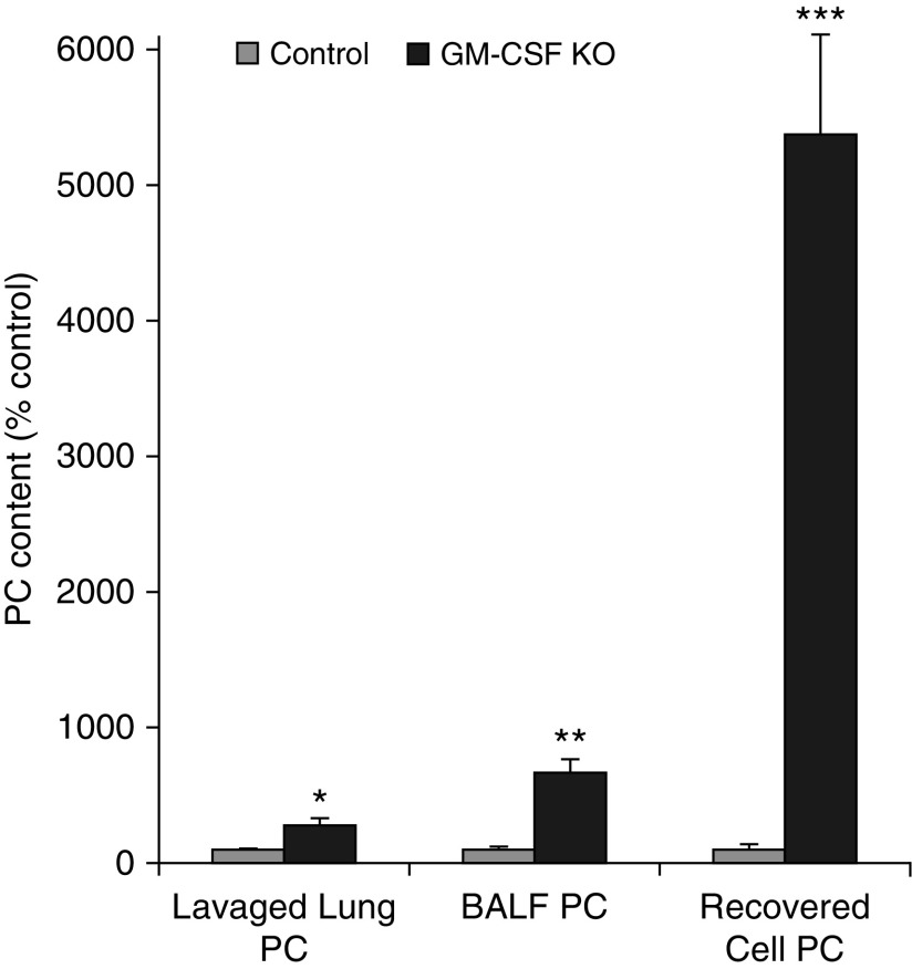
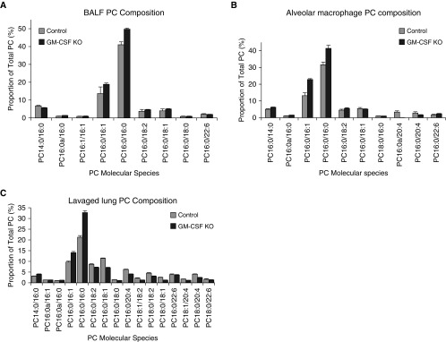
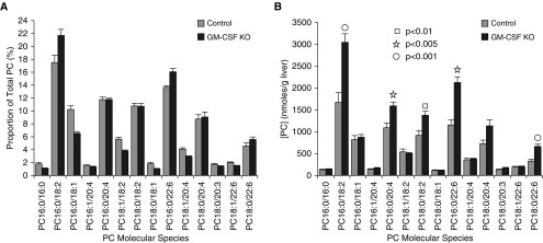
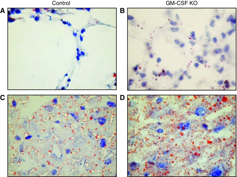
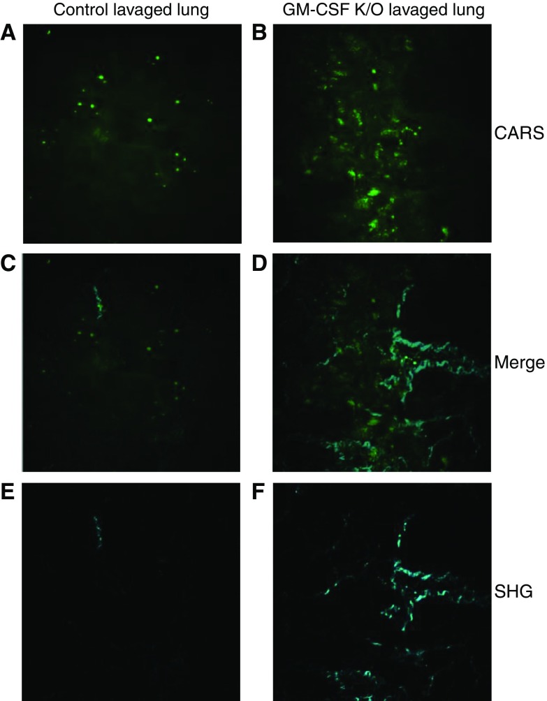
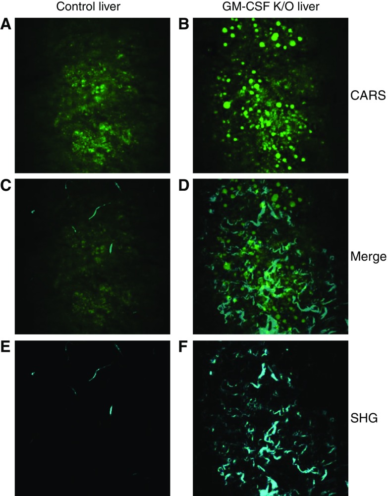
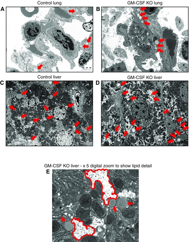
Maintenance of tissue-specific organ lipid compositions characterizes mammalian lipid homeostasis. The lungs and liver synthesize mixed phosphatidylcholine (PC) molecular species that are subsequently tailored for function. The lungs progressively enrich disaturated PC directed to lamellar body surfactant stores before secretion. The liver accumulates polyunsaturated PC directed to very-low-density lipoprotein assembly and secretion, or to triglyceride stores. In each tissue, selective PC species enrichment mechanisms lie at the heart of effective homeostasis. We tested for potential coordination between these spatially separated but possibly complementary phenomena under a major derangement of lung PC metabolism, pulmonary alveolar proteinosis (PAP), which overwhelms homeostasis and leads to excessive surfactant accumulation. Using static and dynamic lipidomics techniques, we compared (1) tissue PC compositions and contents, and (2) in lungs, the absolute rates of synthesis in both control mice and the granulocyte-macrophage colony-stimulating factor knockout model of PAP. Significant disaturated PC accumulation in bronchoalveolar lavage fluid, alveolar macrophage, and lavaged lung tissue occurred alongside increased PC synthesis, consistent with reported defects in alveolar macrophage surfactant turnover. However, microscopy using oil red O staining, coherent anti-Stokes Raman scattering, second harmonic generation, and transmission electron microscopy also revealed neutral-lipid droplet accumulations in alveolar lipofibroblasts of granular macrophage colony-stimulating factor knockout animals, suggesting that lipid homeostasis deficits extend beyond alveolar macrophages. PAP plasma PC composition was significantly polyunsaturated fatty acid enriched, but the content was unchanged and hepatic polyunsaturated fatty acid-enriched PC content increased by 50% with an accompanying micro/macrovesicular steatosis and a fibrotic damage pattern consistent with nonalcoholic fatty liver disease. These data suggest a hepatopulmonary axis of PC metabolism coordination, with wider implications for understanding and managing lipid pathologies in which compromise of one organ has unexpected consequences for another.

Keywords: fibrotic damage; hepatic steatosis; lipidomics; lipotoxicity; pulmonary alveolar proteinosis.


Figures

Figure 1.
Figure 1.
Phosphatidylcholine (PC) accumulations in lung compartments. The mean PC content of control samples were defined as 100% ± SEM while increased content of equivalent samples from granulocyte–macrophage colony-stimulating factor–ablated animals expressed as a percentage of relevant control mean ± SEM. Lavaged lung, n = 12 and 24; bronchoalveolar lavage fluid (BALF), n = 9 and 19; recovered cells, n = 11 and 14. *P < 0.02, **P < 0.0003, ***P < 0.000002. GM-CSF, granulocyte–macrophage colony-stimulating factor knockout; KO, knockout.
Figure 2.
Figure 2.
PC molecular species compositions in lung compartments. (A) Proportional representation of all PC species > 0.5% total PC in lavaged lung tissue from control and GM-CSF KO animals (mean ± SEM, n = 12 and 24). (B) Proportional representation of all PC species > 0.5% total PC in BALF from control and GM-CSF KO animals (mean ± SEM, n = 9 and 19). (C) Proportional representation of all PC species > 0.5% total PC in cells recovered from control and GM-CSF BALF (mean ± SEM, n = 11 and 14). The reported molecular species represent the experimentally ascertained dominant molecular species from the potential isobaric options.
Figure 3.
Figure 3.
PC molecular species compositions and contents in whole liver. (A) Proportional representation of all PC species > 0.5% total PC in whole liver from control and GM-CSF KO animals (mean ± SEM, n = 8 and 20) consistent with relative enrichment of polyunsaturated PC. (B) Contents of the same molecular species per gram wet weight liver. With the exception of PC16:0/18:2, all PC species that were elevated significantly in GM-CSF livers as shown were polyunsaturated.
Figure 4.
Figure 4.
Oil red O (ORO) staining of lavaged lung sections and liver sections. Lavaged lung and liver samples were stained with ORO, counterstained with Mayer’s hematoxylin, and observed using a ×100 oil immersion lens. (A) Representative control lavaged lung section with sporadic ORO-stained inclusions. (B) Representative GM-CSF lavaged lung with many more ORO-stained inclusions, apparently in alveolar type 1 (AT1) respiratory epithelia. (C) Representative control liver section with ORO-stained droplets distributed largely in the sinusoids. (D) Representative GM-CSF liver section with widespread ORO-stained lipid droplet inclusions of varying size within hepatocytes as well as sinusoids, a pattern consistent with steatosis.
Figure 5.
Figure 5.
Coherent anti-Stokes Raman scattering (CARS) and second harmonic generation (SHG) of lung sections. Lavaged lungs from control and GM-CSF KO animals were subjected to CARS and SHG microscopy. A and B show CARS images that confirm the significant increases in lipid droplets present in GM-CSF lungs. C and D show the respective overlays of images A with E, and B with F and define the spatial relationships between lipid accretion and fibrotic changes. E and F show the SHG images of the same areas, which show a pattern of increased collagen fibers in the GM-CSF KO lungs.
Figure 6.
Figure 6.
CARS and SHG of liver sections. Livers from control and GM-CSF KO animals were subjected to CARS and SHG microscopy. A and B show CARS images which confirm significant increases in number and size of lipid droplets present in GM-CSF livers. C and D show the respective overlays of images A with E, and B with F and define the spatial relationships between lipid accretion and hepatic fibrotic changes. E and F show the SHG images of the same areas which show a pattern of significantly increased collagen fiber depositions in the GM-CSF KO livers.
Figure 7.
Figure 7.
Transmission electron microscopy (TEM) of lung and liver tissues. Unlavaged lungs and livers from control and GM-CSF KO animals were subjected to TEM. For control lungs, 12 distinct image fields were collected and analyzed from three grids. For GM-CSF KO lungs, 17 distinct image fields were collected and analyzed from four grids. For control livers, 9 distinct image fields were collected and analyzed from 3 grids. For GM-CSF KO livers 12 distinct image fields were collected and analyzed from 4 grids. Scale bars are 2,000 nm and 5,000 nm for lung tissue and liver tissue, respectively. (A) Control lung: representative image showing a few sporadic lipid droplets (indicated by red arrows) but no consistent pattern. (B) Representative image showing accumulations of lipid droplets largely confined to lipofibroblast cells, with no indication of involvement of the adjacent AT2 or AT1 cells. A larger version of B is presented in Figure E5, which shows more detailed labeling of cells and lipid contents. (C) Control liver: representative image showing the presence of a number of lipid droplets (red arrows), some of which consist of nascent lipoproteins and others that are more likely lipid stores. (D) GM-CSF KO liver, representative image showing a much wider range of lipid droplets (red arrows), which in addition to lipoproteins reveal, when zoomed in upon in E, clusters of very small droplets (outlined in red) alongside larger droplets bounded by lipid bilayers.

References

    1. Jackowski S. Cell cycle regulation of membrane phospholipid metabolism. J Biol Chem. 1996;271:20219–20222. - PubMed
    1. Unger RH, Clark GO, Scherer PE, Orci L. Lipid homeostasis, lipotoxicity and the metabolic syndrome. Biochim Biophys Acta. 2010;1801:209–214. - PubMed
    1. Szendroedi J, Roden M. Ectopic lipids and organ function. Curr Opin Lipidol. 2009;20:50–56. - PubMed
    1. Haffar T, Bérubé-Simard F, Bousette N. Impaired fatty acid oxidation as a cause for lipotoxicity in cardiomyocytes. Biochem Biophys Res Commun. 2015;468:73–78. - PubMed
    1. Guebre-Egziabher F, Alix PM, Koppe L, Pelletier CC, Kalbacher E, Fouque D, Soulage CO. Ectopic lipid accumulation: A potential cause for metabolic disturbances and a contributor to the alteration of kidney function. Biochimie. 2013;95:1971–1979. - PubMed

Publication types

Substances