. 2017 Jun 20;8(25):41364-41378.
doi: 10.18632/oncotarget.17326.

Oleate-induced PTX3 promotes head and neck squamous cell carcinoma metastasis through the up-regulation of vimentin

Affiliations

Oleate-induced PTX3 promotes head and neck squamous cell carcinoma metastasis through the up-regulation of vimentin

Shih-Hung Chan et al. Oncotarget. .

Abstract

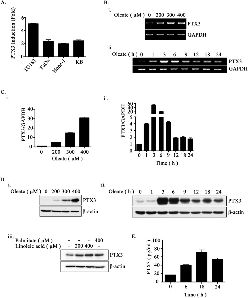
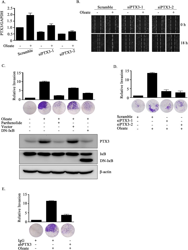
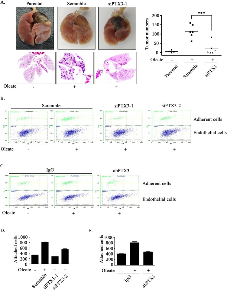
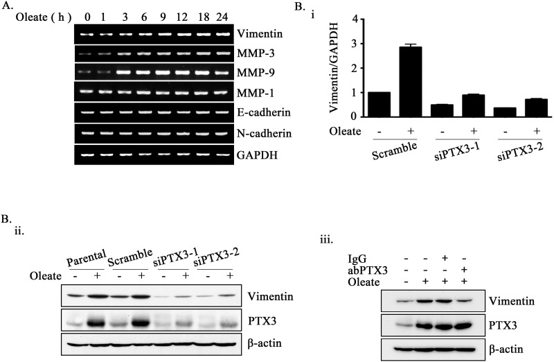
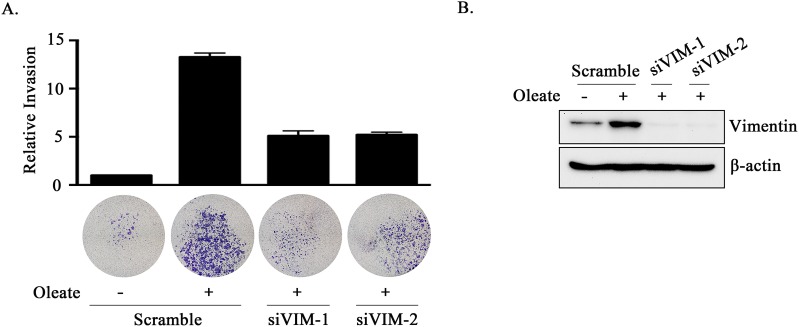
The association between metabolic diseases and the risk of developing cancer is emerging. However, the impact of long pentraxin-3 (PTX3) on dyslipidemia-associated tumor metastasis remains unknown. In this study, we found that oleate induced PTX3 expression and secretion through the activation of Akt/NF-κB pathway in head and neck squamous cell carcinomas (HNSCCs). The activation of NF-κB was essential for the oleate-induced stabilization of PTX3 mRNA. In addition, both the depletion of PTX3 and the inhibition of NF-κB significantly inhibited oleate-induced tumor cell migration and invasion. The enhancement of binding between tumor and endothelial cells was observed in oleate-treated cells but not in the depletion and neutralization of PTX3 with siPTX3 and anti-PTX3 antibodies, respectively. The levels of oleate-induced epithelial-mesenchymal transition (EMT) markers, such as vimentin and MMP-3, were significantly reduced in PTX3-depleted cells. Knocking down vimentin also repressed oleate-induced HNSCC invasion. Furthermore, the depletion of PTX3 blocked the oleate-primed metastatic seeding of tumor cells in the lungs. These results demonstrate that oleate enhances HNSCC metastasis through the PTX3/vimentin signaling axes. The inhibition of PTX3 could be a potential strategy for the treatment of dyslipidemia-mediated HNSCC metastasis.

Keywords: PTX3; metastasis; oleate.


Conflict of interest statement

CONFLICTS OF INTEREST

The authors declare no conflicts of interest.

Figures

Figure 1
Figure 1. Oleate induces the expression of PTX3 in HNSCC cells
(A) The head and neck cancer cell lines TU183, KB, FaDu and HONE1 were treated with 200 μM oleate for 6 h. The mRNA expression of PTX3 was normalized to the GAPDH mRNA level by real-time quantitative PCR. (B-D) TU183 cells were treated with varying concentrations of oleate, linoleic acid and palmitate for 6 h or varying periods of time as indicated. The expression levels of PTX3 mRNA were analyzed by RT-PCR (B) and quantitative PCR (C). The protein levels in cell lysates were determined by Western blotting with antibodies against PTX3 and β-actin (D). (E) The conditioned media from 400 μM oleate-treated TU183 cells was collected to analyze the protein levels of PTX3 by ELISA. The values represent the mean ± s.e.m. of three determinations.
Figure 2
Figure 2. The activation of NF-κB is essential for oleate-induced PTX3 expression
(A and B) TU183 cells were treated with 400 μM oleate for the indicated period of time. The protein levels were determined by Western blotting with antibodies against AKT, IκBα, β-actin and phosphorylated AKT and IκBα. The expression of IkBα was calculated by image-based computational quantification (A). The cytoplasmic fractions and nuclear extracts of the cells were prepared in the same volume of buffer, and an aliquot of each fraction was used for Western blotting analysis using antibodies against NF-κB, HDAC-1 and α-Tubulin (B). (C) Cells were pretreated with 20 μM LY294002 (Sigma-Aldrich, St Louis, MO, USA) for 1 h, then with 400 μM oleate for 1 h (i) or 6 h (ii). The protein levels in cell lysates were determined by Western blotting with antibodies against AKT, PTX3, phosphorylated AKT and β-actin. (D and E) Cells were transfected with the DN-IκB expression vector by lipofection or pretreated with 10 μM parthenolide (Sigma-Aldrich, St Louis, MO, USA) or 5 μM U0126 (Sigma-Aldrich, St Louis, MO, USA) for 1 h, then with 400 μM oleate for 1 h (i) or 6 h (ii) (D). The protein levels in cell lysates were determined by Western blotting with antibodies against ERK, PTX3, IκBα, β-actin and phosphorylated IκBα and ERK. (F and G) Cells were treated with 400 μM oleate for 1 h and then treated with 4 μM actinomycin D (Sigma-Aldrich, St Louis, MO, USA) for the indicated period of time (F). On the other hand, cells were transfected with the DN-IκB expression vector by lipofection or treated with 10 μM parthenolide and 400 μM oleate for 1 h, and then treated with 4 μM actinomycin D for 3 h (G). The expression of PTX3 mRNA was analyzed by real-time quantitative PCR. Relative levels of PTX3 were normalized by GAPDH and the degradation rate was calculated by comparing to 0 h (considered as 100%). The values represent the mean ± s.e.m. of three determinations.
Figure 3
Figure 3. The depletion of PTX3 inhibits oleate-induced HNSCC migration and invasion
(A) TU183 cells were transfected with 20 nM PTX3 siRNA (siPTX3) or scrambled oligonucleotides and treated with or without 400 μM oleate for 18 h. The mRNA expression levels of PTX3 was normalized to the GAPDH mRNA level by real-time quantitative PCR. (B and D) TU183 cells were transfected with 20 nM PTX3 siRNA (siPTX3) or scrambled oligonucleotides by lipofection and then treated with 400 μM oleate for 18 h and 72 h for the migration and invasion assays, respectively. The wound-healing assay was performed as described in the “Materials and Methods” section. The migrating cells were examined using a microscope (B). The invasive properties of the cells were examined using an invasion assay as described in the “Materials and Methods” section. The invading cells were fixed and stained with crystal violet and then examined using a microscope or the cells were solubilized with acetic acid, and the absorbance (OD, 595 nm) was measured in a microplate reader. The values are displayed the mean ± s.e.m. (C-E) TU183 cells were transfected with the DN-IκB expression vector by lipofection or treated with 10 μM parthenolide and then with 400 μM oleate (C), immunoglobulin (IgG) or anti-PTX3 antibodies (1 μg/ml) (E). The invasive properties of the cells were examined and measured. The values are the mean ± s.e.m.
Figure 4
Figure 4. Oleate-induced autocrine production of PTX3 enhances tumor metastasis
(A) TU183 cells were transfected with 20 nM PTX3 siRNA (siPTX3) or scrambled oligonucleotides by lipofection. A lung-colonization analysis was performed by injecting 1 × 10⁶ TU183 cells into the lateral tail vein of SCID mice. Prior to the injection, oleate was injected into the tail vein of mice to mimic the condition of patients who present with 400 μM circulating FFAs. Lung micronodules were examined and photographed after the mice were sacrificed at 6 weeks. The lungs and tumor tissues stained with H&E were examined under a microscope (left panel). The number of micronodules was counted under a microscope (right panel). Parental indicates TU183 cells, either with (N = 6) or without (N = 4) treatment with oleate. siPTX3 (siPTX3-1: N = 3, siPTX3-2: N = 3) indicates the knockdown of PTX3. The values represent the mean ± s.e.m. ***P <0.001. SC: scrambled oligonucleotides. (B-E) TU183 cells were transfected with 20 nM PTX3 siRNA oligonucleotides (siPTX3) and scrambled siRNA (SC) by lipofection, and the cells were treated with 400 μM oleate or anti-PTX3 antibodies (abPTX3) for 18 h. The cells were then labelled with CFSE and cultured with endothelial cells for 30 min. The bound tumor cells (adherent cells) were analyzed using a flow cytometer. TU183 cells were CFSE-positive, and endothelial cells were CFSE-negative. The bound tumor cells were quantified in three independent experiments by flow cytometry. The values are the mean ± s.e.m.
Figure 5
Figure 5. Oleate-induced PTX3 regulates the expression of vimentin
(A) TU183 cells were treated with 400 μM oleate for the indicated period of time. The mRNA expression levels of EMT markers were examined using RT-PCR. (B) TU183 cells were transfected with 20 nM PTX3 siRNA (siPTX3) or scrambled oligonucleotides and treated with or without 400 μM oleate for 18 h. The mRNA expression levels of vimentin was normalized to the GAPDH mRNA level by real-time quantitative PCR (i). The values represent the mean ± s.e.m. of three determinations. Lysates of cells treated with oleate for 24 h or anti-PTX3 antibodies (abPTX3) (1 μg/ml) were prepared and subjected to SDS-PAGE and analyzed by Western blotting with antibodies against PTX3, vimentin and β-actin (ii and iii).
Figure 6
Figure 6. The knockdown of vimentin inhibits oleate-induced tumor cell invasion
(A and B) TU183 cells were transfected with 20 nM vimentin siRNA (siVIM) or scrambled oligonucleotides by lipofection. The invasive properties of the cells were examined using an invasion assay as described in the “Materials and Methods” section. The invading cells were fixed and stained with crystal violet and then examined using a microscope or the cells were solubilized with acetic acid, and the absorbance (OD, 595 nm) was measured in a microplate reader (A). The values are the mean ± s.e.m.. The expression of vimentin and β-actin was determined by Western blotting (B).

Similar articles

Cited by

References

    1. Forastiere A, Koch W, Trotti A, Sidransky D. Head and neck cancer. N Engl J Med. 2001;345:1890–1900. - PubMed
    1. Carvalho AL, Nishimoto IN, Califano JA, Kowalski LP. Trends in incidence and prognosis for head and neck cancer in the United States: a site-specific analysis of the SEER database. Int J Cancer. 2005;114:806–816. - PubMed
    1. Fidler IJ. The pathogenesis of cancer metastasis: the 'seed and soil' hypothesis revisited. Nat Rev Cancer. 2003;3:453–458. - PubMed
    1. Hursting SD, Hursting MJ. Growth signals, inflammation, and vascular perturbations: mechanistic links between obesity, metabolic syndrome, and cancer. Arterioscler Thromb Vasc Biol. 2012;32:1766–1770. - PubMed
    1. Braun S, Bitton-Worms K, LeRoith D. The link between the metabolic syndrome and cancer. Int J Biol Sci. 2011;7:1003–1015. - PMC - PubMed

MeSH terms